• 文献检索
  • 文档翻译
  • 深度研究
  • 学术资讯
  • Suppr Zotero 插件Zotero 插件
  • 邀请有礼
  • 套餐&价格
  • 历史记录
应用&插件
Suppr Zotero 插件Zotero 插件浏览器插件Mac 客户端Windows 客户端微信小程序
定价
高级版会员购买积分包购买API积分包
服务
文献检索文档翻译深度研究API 文档MCP 服务
关于我们
关于 Suppr公司介绍联系我们用户协议隐私条款
关注我们

Suppr 超能文献

核心技术专利:CN118964589B侵权必究
粤ICP备2023148730 号-1Suppr @ 2026

文献检索

告别复杂PubMed语法,用中文像聊天一样搜索,搜遍4000万医学文献。AI智能推荐,让科研检索更轻松。

立即免费搜索

文件翻译

保留排版,准确专业,支持PDF/Word/PPT等文件格式,支持 12+语言互译。

免费翻译文档

深度研究

AI帮你快速写综述,25分钟生成高质量综述,智能提取关键信息,辅助科研写作。

立即免费体验

人肺间充质干细胞与成纤维细胞的比较蛋白质组学分析。

Comparative proteomic analysis of human lung telocytes with fibroblasts.

作者信息

Zheng Yonghua, Cretoiu Dragos, Yan Guoquan, Cretoiu Sanda Maria, Popescu Laurentiu M, Wang Xiangdong

机构信息

Department of Respirology, Xinhua Hospital Affiliated to Shanghai Jiao Tong University School of Medicine, Shanghai, China.

出版信息

J Cell Mol Med. 2014 Apr;18(4):568-89. doi: 10.1111/jcmm.12290. Epub 2014 Mar 28.

DOI:10.1111/jcmm.12290
PMID:24674459
原文链接:https://pmc.ncbi.nlm.nih.gov/articles/PMC4000110/
Abstract

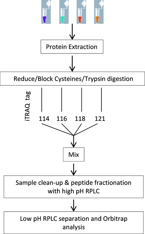
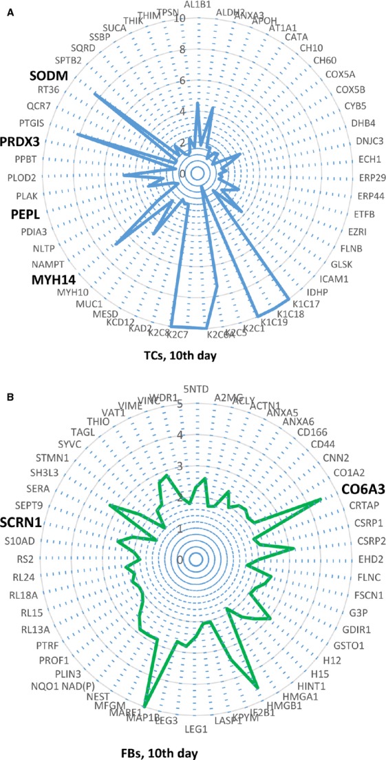
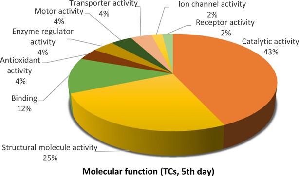
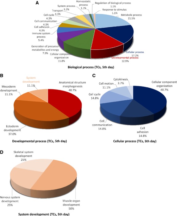
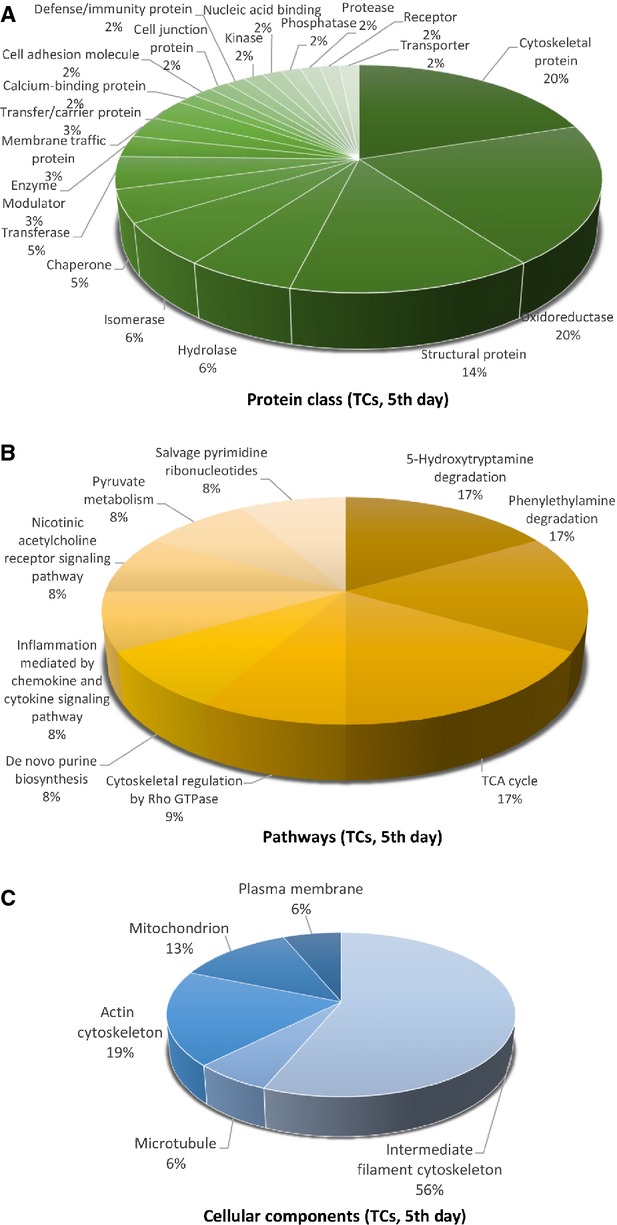
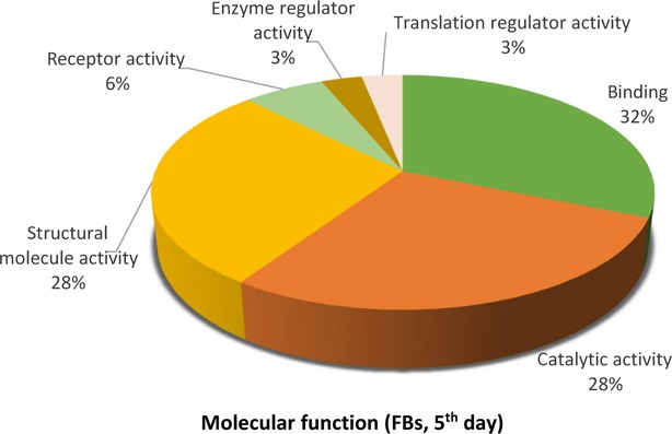
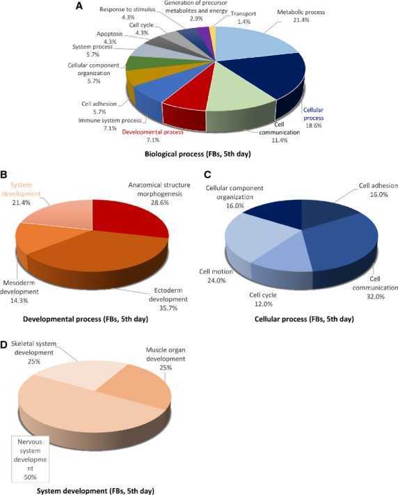
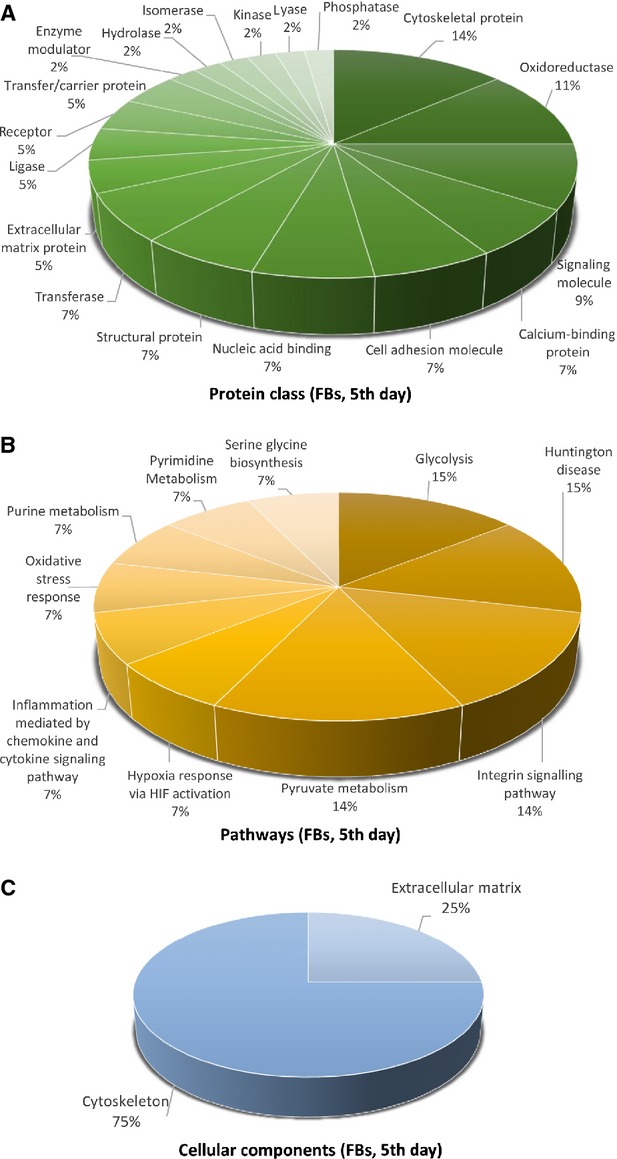
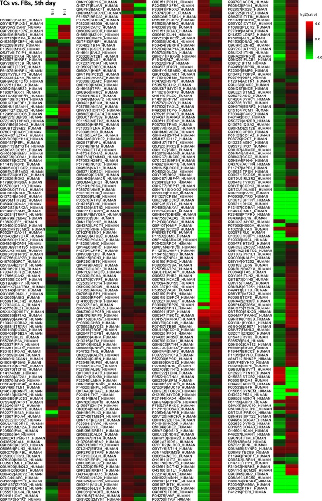
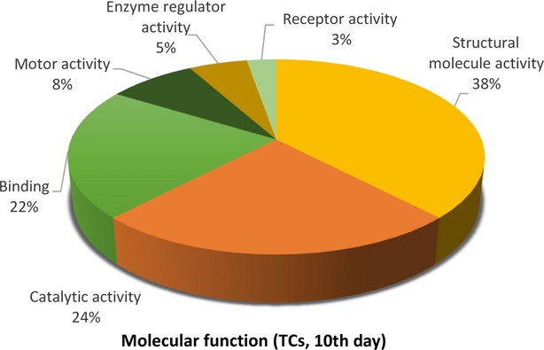
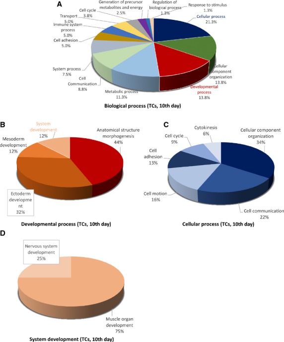
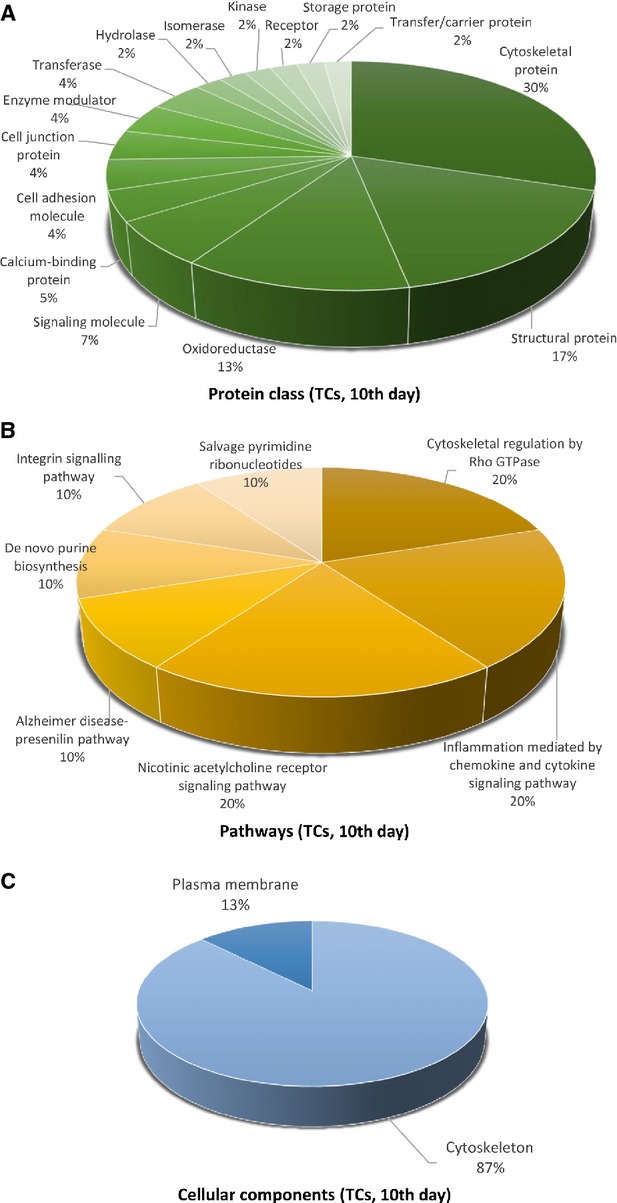
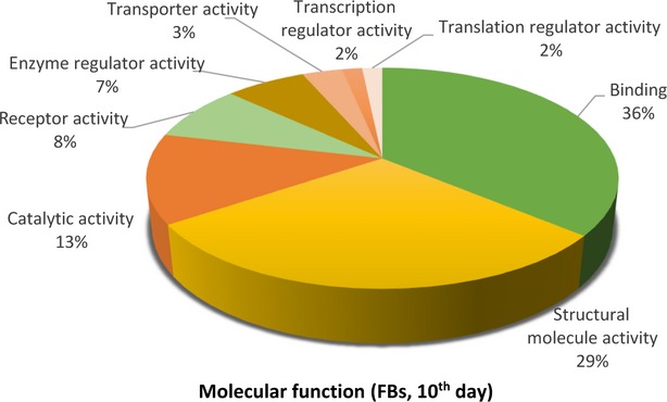
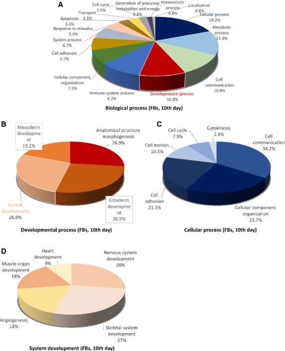
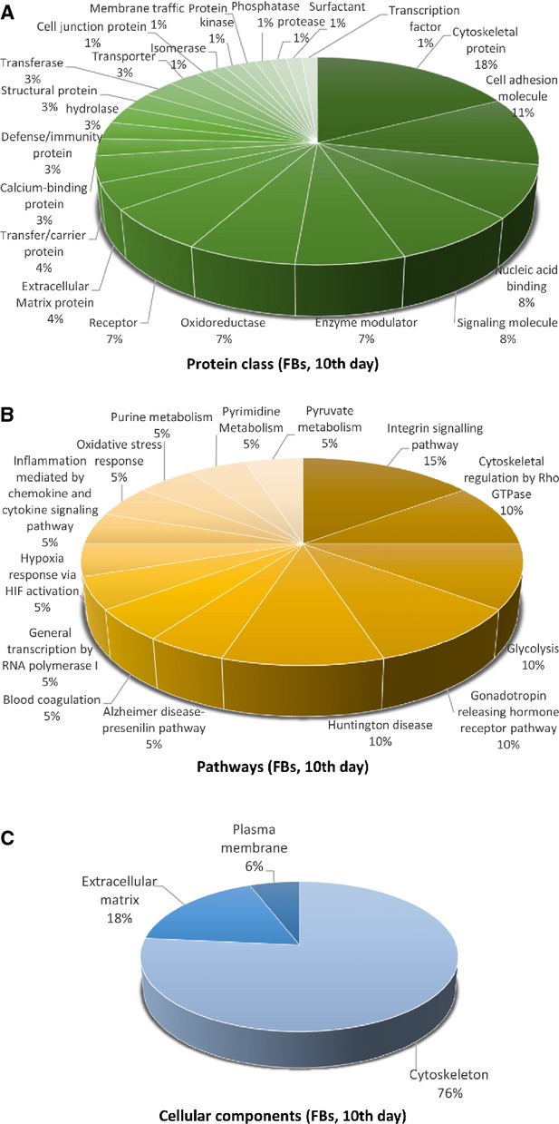
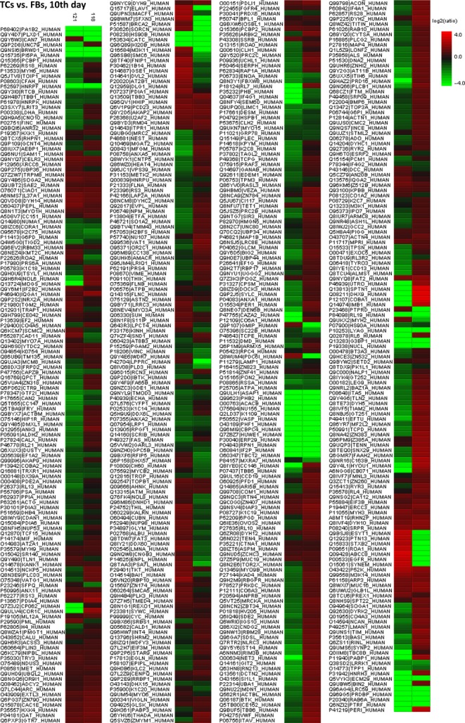
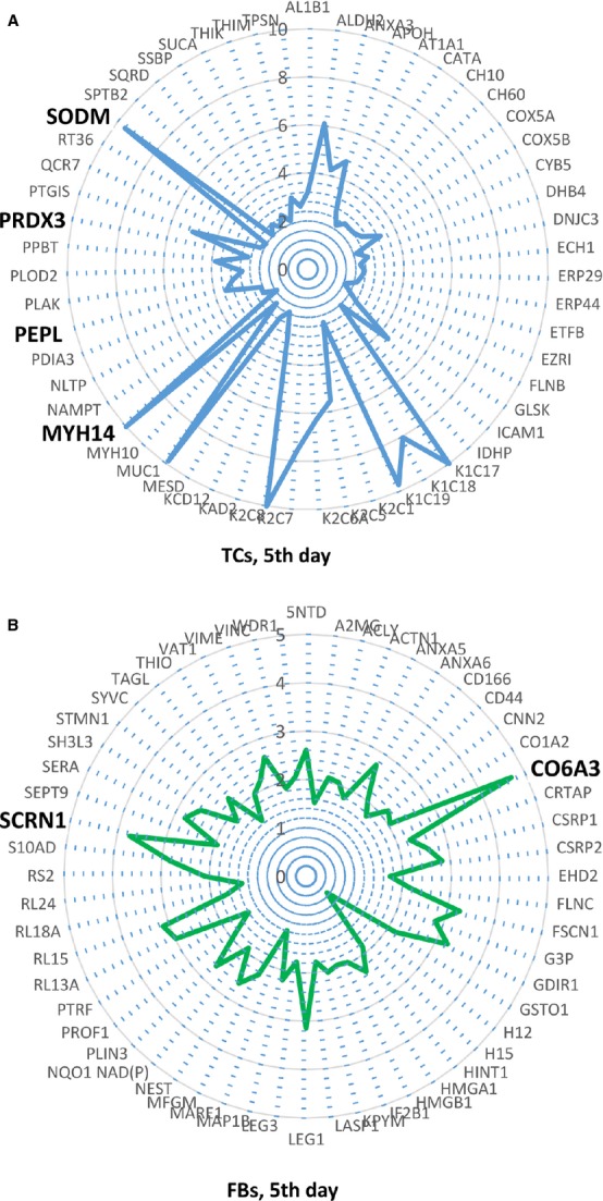
Telocytes (TCs) were recently described as interstitial cells with very long prolongations named telopodes (Tps; www.telocytes.com). Establishing the TC proteome is a priority to show that TCs are a distinct type of cells. Therefore, we examined the molecular aspects of lung TCs by comparison with fibroblasts (FBs). Proteins extracted from primary cultures of these cells were analysed by automated 2-dimensional nano-electrospray ionization liquid chromatography tandem mass spectrometry (2D Nano-ESI LC-MS/MS). Differentially expressed proteins were screened by two-sample t-test (P < 0.05) and fold change (>2), based on the bioinformatics analysis. We identified hundreds of proteins up- or down-regulated, respectively, in TCs as compared with FBs. TC proteins with known identities are localized in the cytoskeleton (87%) and plasma membrane (13%), while FB up-regulated proteins are in the cytoskeleton (75%) and destined to extracellular matrix (25%). These identified proteins were classified into different categories based on their molecular functions and biological processes. While the proteins identified in TCs are mainly involved in catalytic activity (43%) and as structural molecular activity (25%), the proteins in FBs are involved in catalytic activity (24%) and in structural molecular activity, particularly synthesis of collagen and other extracellular matrix components (25%). Anyway, our data show that TCs are completely different from FBs. In conclusion, we report here the first extensive identification of proteins from TCs using a quantitative proteomics approach. Protein expression profile shows many up-regulated proteins e.g. myosin-14, periplakin, suggesting that TCs might play specific roles in mechanical sensing and mechanochemical conversion task, tissue homoeostasis and remodelling/renewal. Furthermore, up-regulated proteins matching those found in extracellular vesicles emphasize TCs roles in intercellular signalling and stem cell niche modulation. The novel proteins identified in TCs will be an important resource for further proteomic research and it will possibly allow biomarker identification for TCs. It also creates the premises for understanding the pathogenesis of some lung diseases involving TCs.

摘要

端粒细胞(TCs)最近被描述为具有名为端粒(Tps;www.telocytes.com)的非常长的延长部分的间质细胞。建立TC蛋白质组是证明TCs是一种独特细胞类型的首要任务。因此,我们通过与成纤维细胞(FBs)比较来研究肺TCs的分子特征。从这些细胞的原代培养物中提取的蛋白质通过自动二维纳米电喷雾电离液相色谱串联质谱法(2D Nano-ESI LC-MS/MS)进行分析。基于生物信息学分析,通过双样本t检验(P < 0.05)和倍数变化(>2)筛选差异表达的蛋白质。与FBs相比,我们分别鉴定出数百种在TCs中上调或下调的蛋白质。具有已知身份的TC蛋白质定位于细胞骨架(87%)和质膜(13%),而FB上调的蛋白质位于细胞骨架(75%)并定位于细胞外基质(25%)。这些鉴定出的蛋白质根据其分子功能和生物学过程被分类为不同类别。虽然在TCs中鉴定出的蛋白质主要参与催化活性(43%)和作为结构分子活性(25%),但FBs中的蛋白质参与催化活性(24%)和结构分子活性,特别是胶原蛋白和其他细胞外基质成分的合成(25%)。无论如何,我们的数据表明TCs与FBs完全不同。总之,我们在此报告首次使用定量蛋白质组学方法对TCs蛋白质进行广泛鉴定。蛋白质表达谱显示许多上调的蛋白质,例如肌球蛋白-14、外周膜蛋白,表明TCs可能在机械传感和机械化学转换任务、组织稳态以及重塑/更新中发挥特定作用。此外,与细胞外囊泡中发现的蛋白质相匹配的上调蛋白质强调了TCs在细胞间信号传导和干细胞微环境调节中的作用。在TCs中鉴定出的新蛋白质将是进一步蛋白质组学研究的重要资源,并且可能允许鉴定TCs的生物标志物。它也为理解一些涉及TCs的肺部疾病的发病机制创造了前提条件。

https://cdn.ncbi.nlm.nih.gov/pmc/blobs/0e57/4000110/ea9354f9cca8/jcmm0018-0568-f17.jpg
https://cdn.ncbi.nlm.nih.gov/pmc/blobs/0e57/4000110/879002466651/jcmm0018-0568-f1.jpg
https://cdn.ncbi.nlm.nih.gov/pmc/blobs/0e57/4000110/5c0fae3a3d0e/jcmm0018-0568-f2.jpg
https://cdn.ncbi.nlm.nih.gov/pmc/blobs/0e57/4000110/54c66027968a/jcmm0018-0568-f3.jpg
https://cdn.ncbi.nlm.nih.gov/pmc/blobs/0e57/4000110/243a47e4a71d/jcmm0018-0568-f4.jpg
https://cdn.ncbi.nlm.nih.gov/pmc/blobs/0e57/4000110/de6a19c944bc/jcmm0018-0568-f5.jpg
https://cdn.ncbi.nlm.nih.gov/pmc/blobs/0e57/4000110/bb629ed22fb6/jcmm0018-0568-f6.jpg
https://cdn.ncbi.nlm.nih.gov/pmc/blobs/0e57/4000110/238e06340b51/jcmm0018-0568-f7.jpg
https://cdn.ncbi.nlm.nih.gov/pmc/blobs/0e57/4000110/efbc5621d083/jcmm0018-0568-f8.jpg
https://cdn.ncbi.nlm.nih.gov/pmc/blobs/0e57/4000110/8586e5ca1842/jcmm0018-0568-f9.jpg
https://cdn.ncbi.nlm.nih.gov/pmc/blobs/0e57/4000110/2762f13194f5/jcmm0018-0568-f10.jpg
https://cdn.ncbi.nlm.nih.gov/pmc/blobs/0e57/4000110/5e0205214466/jcmm0018-0568-f11.jpg
https://cdn.ncbi.nlm.nih.gov/pmc/blobs/0e57/4000110/ff808e8b6642/jcmm0018-0568-f12.jpg
https://cdn.ncbi.nlm.nih.gov/pmc/blobs/0e57/4000110/4c2badfe0275/jcmm0018-0568-f13.jpg
https://cdn.ncbi.nlm.nih.gov/pmc/blobs/0e57/4000110/758cd7470686/jcmm0018-0568-f14.jpg
https://cdn.ncbi.nlm.nih.gov/pmc/blobs/0e57/4000110/88e83efcfd13/jcmm0018-0568-f15.jpg
https://cdn.ncbi.nlm.nih.gov/pmc/blobs/0e57/4000110/f6d89fbcdaaf/jcmm0018-0568-f16.jpg
https://cdn.ncbi.nlm.nih.gov/pmc/blobs/0e57/4000110/ea9354f9cca8/jcmm0018-0568-f17.jpg

相似文献

1
Comparative proteomic analysis of human lung telocytes with fibroblasts.人肺间充质干细胞与成纤维细胞的比较蛋白质组学分析。
J Cell Mol Med. 2014 Apr;18(4):568-89. doi: 10.1111/jcmm.12290. Epub 2014 Mar 28.
2
Protein profiling of human lung telocytes and microvascular endothelial cells using iTRAQ quantitative proteomics.使用iTRAQ定量蛋白质组学对人肺间质细胞和微血管内皮细胞进行蛋白质谱分析。
J Cell Mol Med. 2014 Jun;18(6):1035-59. doi: 10.1111/jcmm.12350.
3
Genetic comparison of mouse lung telocytes with mesenchymal stem cells and fibroblasts.小鼠肺脏 telocytes 与间充质干细胞和成纤维细胞的遗传学比较。
J Cell Mol Med. 2013 Apr;17(4):567-77. doi: 10.1111/jcmm.12052.
4
Differences in the expression of chromosome 1 genes between lung telocytes and other cells: mesenchymal stem cells, fibroblasts, alveolar type II cells, airway epithelial cells and lymphocytes.肺间充质干细胞与其他细胞(间充质干细胞、成纤维细胞、II型肺泡细胞、气道上皮细胞和淋巴细胞)之间1号染色体基因表达的差异。
J Cell Mol Med. 2014 May;18(5):801-10. doi: 10.1111/jcmm.12302.
5
Global analyses of Chromosome 17 and 18 genes of lung telocytes compared with mesenchymal stem cells, fibroblasts, alveolar type II cells, airway epithelial cells, and lymphocytes.与间充质干细胞、成纤维细胞、II型肺泡细胞、气道上皮细胞和淋巴细胞相比,对肺间质细胞17号和18号染色体基因的全局分析。
Biol Direct. 2015 Mar 11;10:9. doi: 10.1186/s13062-015-0042-0.
6
Comparison of Chromosome 4 gene expression profile between lung telocytes and other local cell types.肺间质细胞与其他局部细胞类型之间4号染色体基因表达谱的比较。
J Cell Mol Med. 2016 Jan;20(1):71-80. doi: 10.1111/jcmm.12746. Epub 2015 Dec 17.
7
The secretome of myocardial telocytes modulates the activity of cardiac stem cells.心肌间质细胞的分泌组调节心脏干细胞的活性。
J Cell Mol Med. 2015 Aug;19(8):1783-94. doi: 10.1111/jcmm.12624. Epub 2015 Jul 14.
8
Variations of chromosomes 2 and 3 gene expression profiles among pulmonary telocytes, pneumocytes, airway cells, mesenchymal stem cells and lymphocytes.肺脏间充质干细胞、肺上皮细胞、气道细胞、间充质干细胞和淋巴细胞中2号和3号染色体基因表达谱的变化。
J Cell Mol Med. 2014 Oct;18(10):2044-60. doi: 10.1111/jcmm.12429. Epub 2014 Oct 2.
9
Skin telocytes versus fibroblasts: two distinct dermal cell populations.皮肤间充质干细胞与成纤维细胞:两种不同的真皮细胞群体。
J Cell Mol Med. 2015 Nov;19(11):2530-9. doi: 10.1111/jcmm.12671. Epub 2015 Sep 28.
10
Uterine Telocytes: A Review of Current Knowledge.子宫间充质干细胞:当前知识综述。
Biol Reprod. 2015 Jul;93(1):10. doi: 10.1095/biolreprod.114.125906. Epub 2015 Feb 18.

引用本文的文献

1
Distribution characteristics and morphological comparison of telocytes in the aortic bulb and myocardium of yak heart.牦牛心脏主动脉球和心肌中特络细胞的分布特征及形态学比较
BMC Vet Res. 2025 Feb 22;21(1):88. doi: 10.1186/s12917-025-04553-x.
2
Telocytes of the male reproductive system: dynamic tissue organizers.男性生殖系统的间充质干细胞:动态组织组织者。
Front Cell Dev Biol. 2024 Oct 14;12:1444156. doi: 10.3389/fcell.2024.1444156. eCollection 2024.
3
Telocytes: current methods of research, challenges and future perspectives.

本文引用的文献

1
Phenotypical and ultrastructural features of Oct4-positive cells in the adult mouse lung.成年小鼠肺中Oct4阳性细胞的表型和超微结构特征。
J Cell Mol Med. 2014 Jul;18(7):1321-33. doi: 10.1111/jcmm.12295. Epub 2014 Jun 3.
2
Intramyocardial transplantation of cardiac telocytes decreases myocardial infarction and improves post-infarcted cardiac function in rats.心脏间充质干细胞的心肌内移植可减少大鼠心肌梗死并改善梗死后的心功能。
J Cell Mol Med. 2014 May;18(5):780-9. doi: 10.1111/jcmm.12259. Epub 2014 Mar 21.
3
New insights into the morphogenic role of stromal cells and their relevance for regenerative medicine. lessons from the heart.
间质细胞:当前的研究方法、挑战与未来展望。
Cell Tissue Res. 2024 May;396(2):141-155. doi: 10.1007/s00441-024-03888-5. Epub 2024 Mar 28.
4
"Stromal cells in prostate cancer pathobiology: friends or foes?"."前列腺癌病理生物学中的基质细胞:是敌是友?"
Br J Cancer. 2023 Apr;128(6):930-939. doi: 10.1038/s41416-022-02085-x. Epub 2022 Dec 8.
5
Telocytes in Fibrosis Diseases: From Current Findings to Future Clinical Perspectives.纤维化疾病中的 Telocytes:从现有发现到未来的临床展望。
Cell Transplant. 2022 Jan-Dec;31:9636897221105252. doi: 10.1177/09636897221105252.
6
Dynamic Involvement of Telocytes in Modulating Multiple Signaling Pathways in Cardiac Cytoarchitecture.心肌细胞外基质中长梭形细胞(telocytes/TCs)的动态参与调控多种信号通路。
Int J Mol Sci. 2022 May 21;23(10):5769. doi: 10.3390/ijms23105769.
7
Telocytes reduce oxidative stress by downregulating DUOX2 expression in inflamed lungs of mice.Telocytes 通过下调 DUOX2 表达减轻小鼠肺部炎症中的氧化应激。
Acta Biochim Biophys Sin (Shanghai). 2022 Apr 25;54(4):574-582. doi: 10.3724/abbs.2022017.
8
Roles of pulmonary telocytes in airway epithelia to benefit experimental acute lung injury through production of telocyte-driven mediators and exosomes.肺 telocytes 在气道上皮中的作用通过产生 telocyte 驱动的介质和外泌体有益于实验性急性肺损伤。
Cell Biol Toxicol. 2023 Apr;39(2):451-465. doi: 10.1007/s10565-021-09670-5. Epub 2022 Jan 3.
9
Mechanisms of interactions between lung-origin telocytes and mesenchymal stem cells to treat experimental acute lung injury.肺源间质细胞与间充质干细胞相互作用治疗实验性急性肺损伤的机制
Clin Transl Med. 2020 Dec;10(8):e231. doi: 10.1002/ctm2.231.
10
Telocytes contribute to aging-related modifications in the prostate.间质细胞参与前列腺与衰老相关的改变。
Sci Rep. 2020 Dec 7;10(1):21392. doi: 10.1038/s41598-020-78532-7.
基质细胞的形态发生作用及其与再生医学的相关性的新见解。来自心脏的经验教训。
J Cell Mol Med. 2014 Mar;18(3):363-70. doi: 10.1111/jcmm.12247. Epub 2014 Feb 18.
4
General statistical framework for quantitative proteomics by stable isotope labeling.基于稳定同位素标记的定量蛋白质组学的通用统计框架。
J Proteome Res. 2014 Mar 7;13(3):1234-47. doi: 10.1021/pr4006958. Epub 2014 Feb 10.
5
Electrophysiology of human cardiac atrial and ventricular telocytes.人心房和心室 telocytes 的电生理学。
J Cell Mol Med. 2014 Feb;18(2):355-62. doi: 10.1111/jcmm.12240.
6
A loss of telocytes accompanies fibrosis of multiple organs in systemic sclerosis.全身性硬皮病多个器官纤维化伴随间质细胞丢失。
J Cell Mol Med. 2014 Feb;18(2):253-62. doi: 10.1111/jcmm.12228.
7
Remodeling of cell-cell junctions in arrhythmogenic cardiomyopathy.致心律失常性心肌病中细胞间连接的重塑
Cell Commun Adhes. 2014 Feb;21(1):13-23. doi: 10.3109/15419061.2013.876016.
8
Distribution and characteristics of telocytes as nurse cells in the architectural organization of engineered heart tissues.工程化心脏组织的建筑组织中作为滋养细胞的 telocytes 的分布和特征。
Sci China Life Sci. 2014 Feb;57(2):241-7. doi: 10.1007/s11427-013-4602-1. Epub 2014 Jan 15.
9
Telocytes, exosomes, gap junctions and the cytoskeleton: the makings of a primitive nervous system?间质细胞、外泌体、缝隙连接与细胞骨架:原始神经系统的构成要素?
Front Cell Neurosci. 2014 Jan 3;7:278. doi: 10.3389/fncel.2013.00278. eCollection 2014.
10
Proteomics of extracellular vesicles: Exosomes and ectosomes.细胞外囊泡的蛋白质组学:外泌体与微泡。
Mass Spectrom Rev. 2015 Jul-Aug;34(4):474-90. doi: 10.1002/mas.21420. Epub 2014 Jan 14.