State Key Laboratory of Ophthalmology, Zhongshan Ophthalmic Center, Sun Yat-sen University, Guangzhou, People's Republic of China.
Int J Nanomedicine. 2013;8:1563-72. doi: 10.2147/IJN.S37635. Epub 2013 Apr 19.
To evaluate the effects of small interference RNA protein kinase C-alpha (siRNA-PKCα) on experimental proliferative vitreoretinopathy (PVR) induced by dispase in mice.
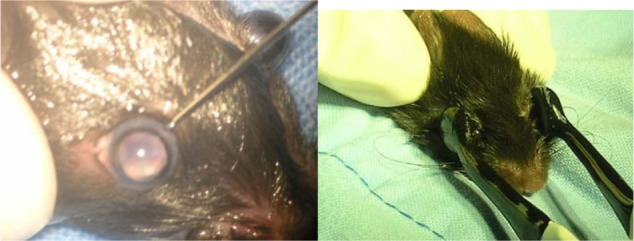
C57BL/6 mice PVR models (4-6 weeks old) were induced by intravitreal injection of dispase and then equally divided into six groups. After 1 week, the five treatment groups received 2 μL, intravitreal injections of siRNA-PKCα at a concentration of 250 nM, 500 nM, 750 nM, 1000 nM, and 1500 nM, respectively, while the negative control group received 2 μL of 500 nM no-silencing siRNA. SiRNA-PKCα was transfected by a square wave electroporator. Postoperative ophthalmic observations of lens clarity and the fundus of the eyes were performed periodically. The eyeballs of the mice were enucleated and imbedded in optimal cutting temperature to perform histological and immunofluorescence analysis at the end of a 4-week observation period.
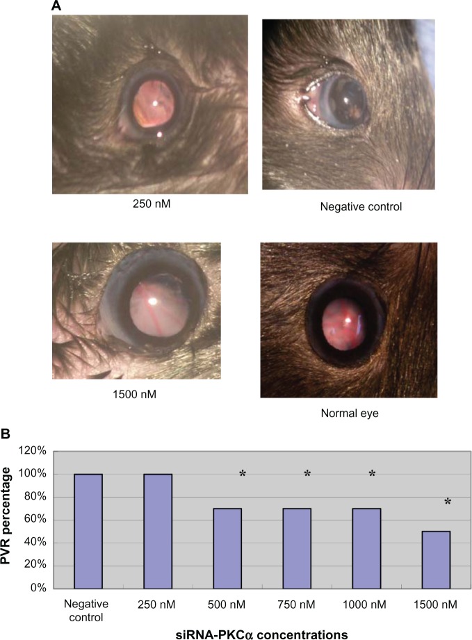
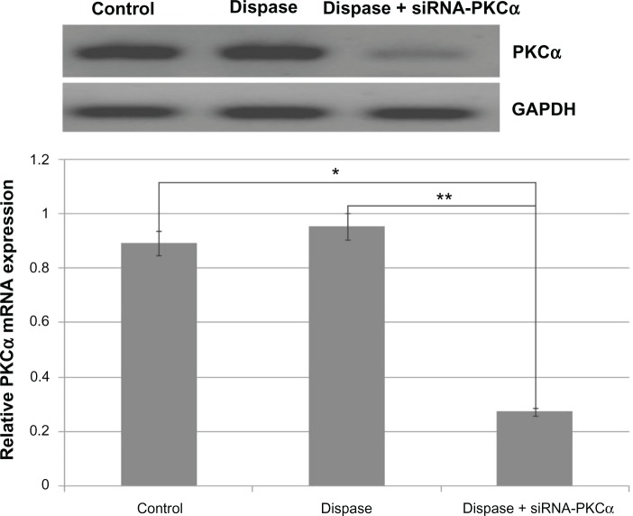
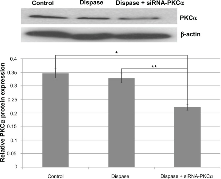
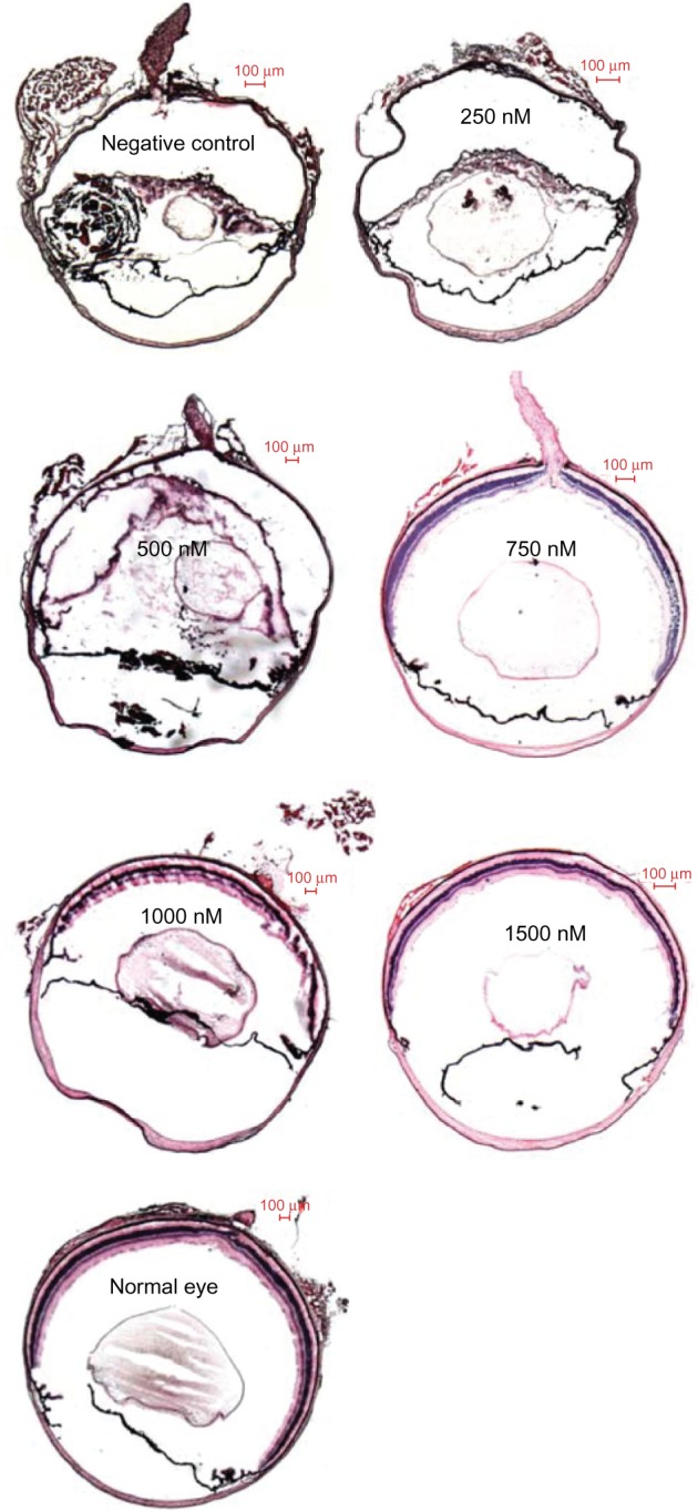
Four weeks after the siRNA-PKCα injections, there are 100% lens dissolution and 100% PVR in the 250 nM group and 70%, 70%, 70%, and 50% PVR in the 500 nM, 750 nM, 1000 nM, and 1500 nM groups, respectively, which is significantly different from the negative group. Abnormalities in fundus appearance were related to the concentrations of siRNA-PKCα; a higher concentration of siRNA-PKCα resulted in a more normal fundus. Histological sections by hematoxylin-eosin staining of the eyes support the clinical observation. Immunofluorescence analysis showed that RPE65, glutamine synthase, glial acidic fibrillary protein, and α-smooth muscle actin were increasing in the retina with the decreasing concentration of siRNA-PKCα, indicating that intraocular siRNA-PKCα can partly inhibit changes of markers for glia cells, fibroblast cells, retinal pigment epithelium cells, and Müller cells in the process of PVR.
Gene therapy with siRNA-PKCα could effectively inhibit PVR in mice and provide us with a novel therapeutic target on PVR.
评价小干扰 RNA 蛋白激酶 C-α(siRNA-PKCα)对Dispase 诱导的实验性增殖性玻璃体视网膜病变(PVR)的作用。
C57BL/6 小鼠 PVR 模型(4-6 周龄)通过玻璃体内注射 Dispase 诱导,然后均等分为 6 组。1 周后,5 个治疗组分别接受 2 μL、浓度为 250 nM、500 nM、750 nM、1000 nM 和 1500 nM 的 siRNA-PKCα 玻璃体腔内注射,阴性对照组接受 2 μL、浓度为 500 nM 的非沉默 siRNA。使用方波电穿孔仪转染 siRNA-PKCα。定期进行晶状体清晰度和眼底的眼部观察。在 4 周观察期结束时,将小鼠眼球取出并包埋在最佳切割温度中,进行组织学和免疫荧光分析。
siRNA-PKCα 注射后 4 周,250 nM 组 100%晶状体溶解和 100%PVR,500 nM、750 nM、1000 nM 和 1500 nM 组分别为 70%、70%、70%和 50%PVR,与阴性组相比差异有统计学意义。眼底外观异常与 siRNA-PKCα 的浓度有关;siRNA-PKCα 浓度越高,眼底越正常。眼组织学切片的苏木精-伊红染色支持临床观察。免疫荧光分析显示,随着 siRNA-PKCα 浓度的降低,RPE65、谷氨酰胺合成酶、胶质酸性纤维蛋白和α-平滑肌肌动蛋白在视网膜中增加,表明眼内 siRNA-PKCα 可部分抑制 PVR 过程中胶质细胞、成纤维细胞、视网膜色素上皮细胞和 Müller 细胞标志物的变化。
siRNA-PKCα 基因治疗可有效抑制小鼠 PVR,为 PVR 提供新的治疗靶点。