Kqiku Lumnije, Biblekaj Robert, Weiglein Andreas H, Kqiku Xhylsime, Städtler Peter
Division of Preventive and Operative Dentistry, Endodontics, Pedodontics, and Minimally Invasive Dentistry, Auenbruggerplatz 4/6, A-8036 Graz, Austria.
Croat Med J. 2013 Apr;54(2):180-4. doi: 10.3325/cmj.2013.54.180.
To describe vascular anatomy of the maxillary sinus in dentate specimens dissected from human cadavers.
Twenty dentate maxillary specimens were dissected, anatomically prepared, and injected with liquid latex for a better visualization of the maxillary sinus artery.
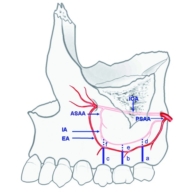
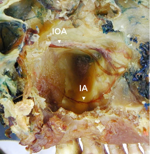
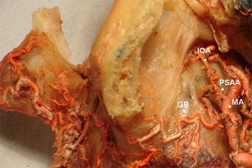
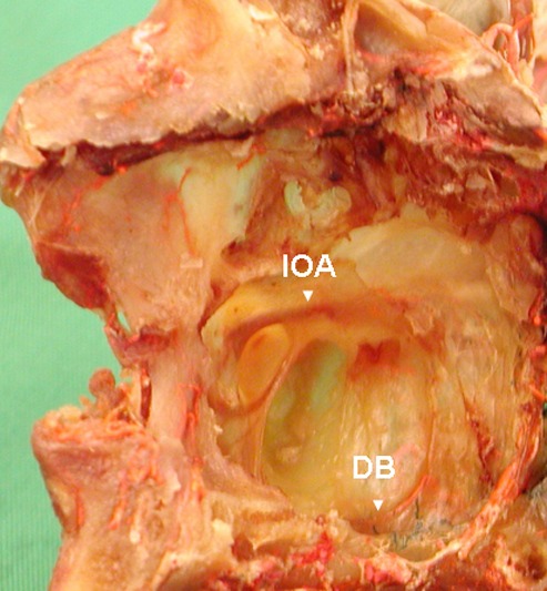
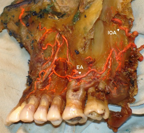
We found an intraosseous anastomosis in 100% and an extraosseous anastomosis in 90% of the cases. The anterior lateral wall of the maxillary sinus was transversed by two anastomoses between the posterior superior alveolar artery (PSAA) and the infraorbital artery (IOA). The PSAA was divided into a gingival and dental branch. The gingival branch anastomosed with the terminal extraosseous branch of the extraosseous anastomosis (EOA) and the dental branch with the intraosseous branch of the intraosseous anastomosis (IOA). The mean distances from the alveolar ridge to the extraosseus anastomosis were 16 mm for the second maxillary molar, 12.3 mm for the first maxillary molar, and 13.1 mm for the second maxillary premolar. The mean distances from the intraosseous anastomosis to the alveolar ridge were 17.7 mm for the second maxillary molar, 14.5 mm for the first maxillary molar, and 14.66 mm for the second maxillary premolar.
These findings provide relevant data for clinical dentistry in order to avoid bleeding complications and minimize the risk of injury to the arterial network of the maxillary sinus during surgical procedures in the dentate maxilla region.
描述从人体尸体上解剖得到的有牙标本中上颌窦的血管解剖结构。
解剖20个有牙的上颌标本,进行解剖学处理,并注入液体乳胶以更好地观察上颌窦动脉。
我们发现100%的病例存在骨内吻合,90%的病例存在骨外吻合。上颌窦前外侧壁有两条后上牙槽动脉(PSAA)与眶下动脉(IOA)之间的吻合支穿过。PSAA分为牙龈支和牙支。牙龈支与骨外吻合(EOA)的终末骨外分支吻合,牙支与骨内吻合(IOA)的骨内分支吻合。从牙槽嵴到骨外吻合的平均距离,上颌第二磨牙为16mm,上颌第一磨牙为12.3mm,上颌第二前磨牙为13.1mm。从骨内吻合到牙槽嵴的平均距离,上颌第二磨牙为17.7mm,上颌第一磨牙为14.5mm,上颌第二前磨牙为14.66mm。
这些发现为临床牙科提供了相关数据,以便在有牙上颌区域的外科手术中避免出血并发症,并将上颌窦动脉网络损伤的风险降至最低。