Department of Animal Biology, School of Veterinary Medicine, University of Pennsylvania, Philadelphia, Pennsylvania, United States of America.
PLoS One. 2013 Apr 22;8(4):e62410. doi: 10.1371/journal.pone.0062410. Print 2013.
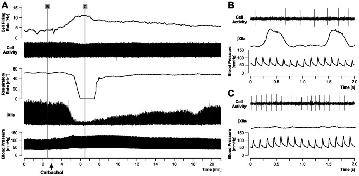
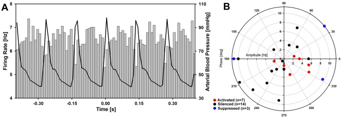
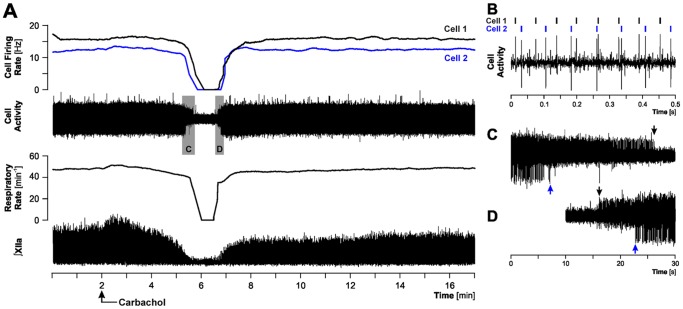
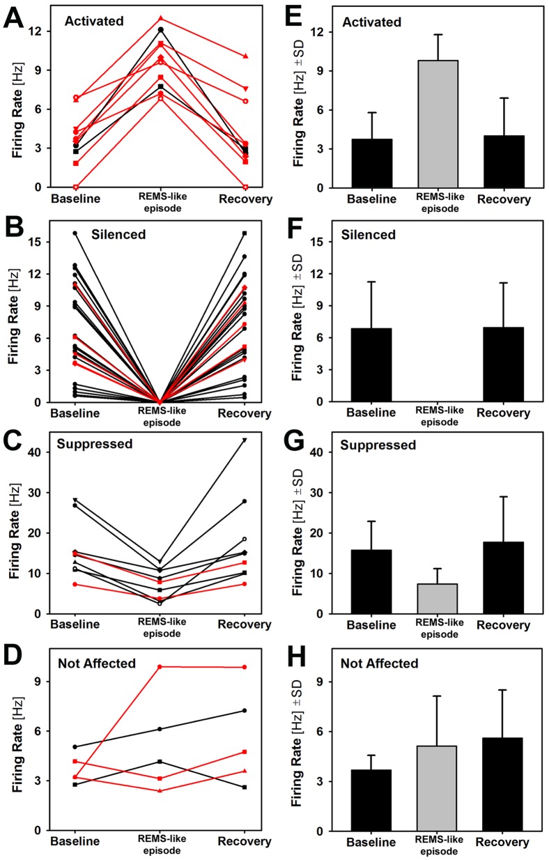
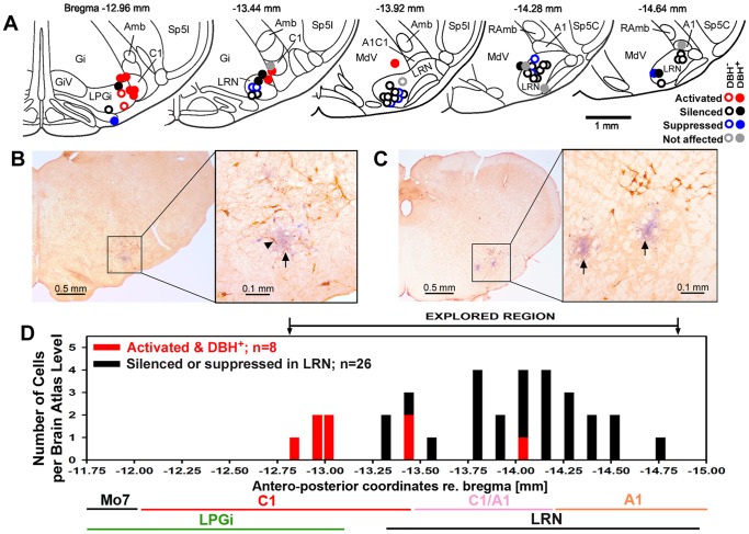
Rapid eye movement sleep (REMS) is generated in the brainstem by a distributed network of neurochemically distinct neurons. In the pons, the main subtypes are cholinergic and glutamatergic REMS-on cells and aminergic REMS-off cells. Pontine REMS-on cells send axons to the ventrolateral medulla (VLM), but little is known about REMS-related activity of VLM cells. In urethane-anesthetized rats, dorsomedial pontine injections of carbachol trigger REMS-like episodes that include cortical and hippocampal activation and suppression of motoneuronal activity; the episodes last 4-8 min and can be elicited repeatedly. We used this model to determine whether VLM catecholaminergic cells are silenced during REMS, as is typical of most aminergic neurons studied to date, and to investigate other REMS-related cells in this region. In 18 anesthetized, paralyzed and artificially ventilated rats, we obtained extracellular recordings from VLM cells when REMS-like episodes were elicited by pontine carbachol injections (10 mM, 10 nl). One major group were the cells that were activated during the episodes (n = 10). Their baseline firing rate of 3.7±2.1 (SD) Hz increased to 9.7±2.1 Hz. Most were found in the adrenergic C1 region and at sites located less than 50 µm from dopamine β-hydroxylase-positive (DBH(+)) neurons. Another major group were the silenced or suppressed cells (n = 35). Most were localized in the lateral reticular nucleus (LRN) and distantly from any DBH(+) cells. Their baseline firing rates were 6.8±4.4 Hz and 15.8±7.1 Hz, respectively, with the activity of the latter reduced to 7.4±3.8 Hz. We conclude that, in contrast to the pontine noradrenergic cells that are silenced during REMS, medullary adrenergic C1 neurons, many of which drive the sympathetic output, are activated. Our data also show that afferent input transmitted to the cerebellum through the LRN is attenuated during REMS. This may distort the spatial representation of body position during REMS.
快速眼动睡眠(REMS)是由分布在不同神经化学神经元网络的脑干产生的。在脑桥上,主要的亚型是胆碱能和谷氨酸能的 REMS-on 细胞和单胺能的 REMS-off 细胞。脑桥上的 REMS-on 细胞将轴突发送到腹外侧髓质(VLM),但关于 VLM 细胞与 REMS 相关的活动知之甚少。在乌拉坦麻醉的大鼠中,内侧脑桥注射卡巴胆碱会引发类似 REMS 的发作,包括皮质和海马的激活以及运动神经元活动的抑制;发作持续 4-8 分钟,可以反复引发。我们使用该模型来确定 VLM 儿茶酚胺能细胞是否在 REMS 期间被沉默,这与迄今为止研究的大多数单胺能神经元典型情况一样,并研究该区域的其他与 REMS 相关的细胞。在 18 只麻醉、瘫痪和人工通气的大鼠中,当脑桥卡巴胆碱注射(10 mM,10 nl)引发类似 REMS 的发作时,我们从 VLM 细胞中获得了细胞外记录。一组主要是在发作期间被激活的细胞(n = 10)。它们的基线放电率为 3.7±2.1(SD)Hz,增加到 9.7±2.1 Hz。大多数位于肾上腺素能 C1 区,并且位于距离多巴胺 β-羟化酶阳性(DBH(+))神经元小于 50 µm 的位置。另一组主要是沉默或抑制的细胞(n = 35)。大多数位于外侧网状核(LRN),并且远离任何 DBH(+)细胞。它们的基线放电率分别为 6.8±4.4 Hz 和 15.8±7.1 Hz,后者的活动减少到 7.4±3.8 Hz。我们得出的结论是,与在 REMS 期间被沉默的脑桥上的去甲肾上腺素能细胞相反,许多驱动交感神经输出的髓质肾上腺素能 C1 神经元被激活。我们的数据还表明,通过 LRN 传递到小脑的传入输入在 REMS 期间被减弱。这可能会在 REMS 期间扭曲身体位置的空间表示。