• 文献检索
  • 文档翻译
  • 深度研究
  • 学术资讯
  • Suppr Zotero 插件Zotero 插件
  • 邀请有礼
  • 套餐&价格
  • 历史记录
应用&插件
Suppr Zotero 插件Zotero 插件浏览器插件Mac 客户端Windows 客户端微信小程序
定价
高级版会员购买积分包购买API积分包
服务
文献检索文档翻译深度研究API 文档MCP 服务
关于我们
关于 Suppr公司介绍联系我们用户协议隐私条款
关注我们

Suppr 超能文献

核心技术专利:CN118964589B侵权必究
粤ICP备2023148730 号-1Suppr @ 2026

文献检索

告别复杂PubMed语法,用中文像聊天一样搜索,搜遍4000万医学文献。AI智能推荐,让科研检索更轻松。

立即免费搜索

文件翻译

保留排版,准确专业,支持PDF/Word/PPT等文件格式,支持 12+语言互译。

免费翻译文档

深度研究

AI帮你快速写综述,25分钟生成高质量综述,智能提取关键信息,辅助科研写作。

立即免费体验

用于中耳和内耳精确成像和空间重建的三维组织学标本制备。

Three-dimensional histological specimen preparation for accurate imaging and spatial reconstruction of the middle and inner ear.

机构信息

Department of Otolaryngology, Institute for AudioNeurotechnology (VIANNA), Hannover Medical School, Carl-Neuberg-Str. 1, 30625, Hannover, Germany.

出版信息

Int J Comput Assist Radiol Surg. 2013 Jul;8(4):481-509. doi: 10.1007/s11548-013-0825-7. Epub 2013 Apr 30.

DOI:10.1007/s11548-013-0825-7
PMID:23633112
原文链接:https://pmc.ncbi.nlm.nih.gov/articles/PMC3702969/
Abstract

PURPOSE

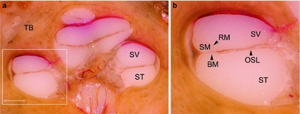
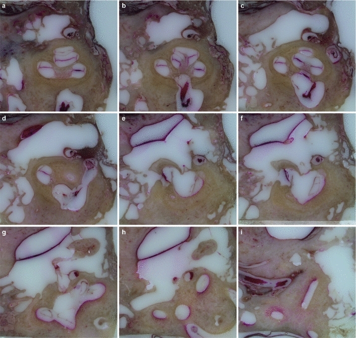
This paper presents a highly accurate cross-sectional preparation technique. The research aim was to develop an adequate imaging modality for both soft and bony tissue structures featuring high contrast and high resolution. Therefore, the advancement of an already existing micro-grinding procedure was pursued. The central objectives were to preserve spatial relations and to ensure the accurate three-dimensional reconstruction of histological sections.

METHODS

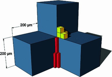
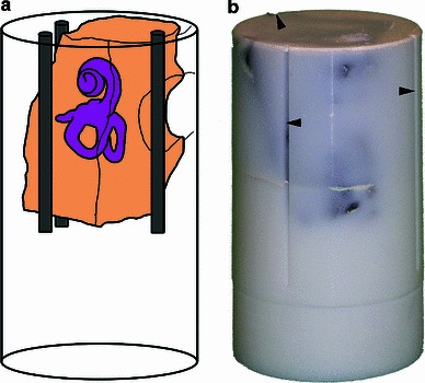
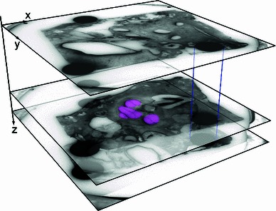
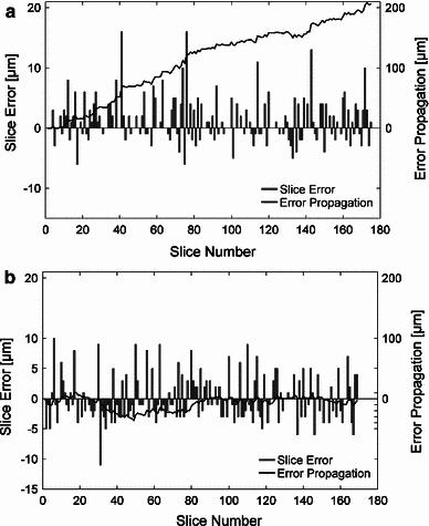
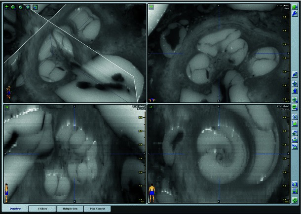
Twelve human temporal bone specimens including middle and inner ear structures were utilized. They were embedded in epoxy resin, then dissected by serial grinding and finally digitalized. The actual abrasion of each grinding slice was measured using a tactile length gauge with an accuracy of one micrometre. The cross-sectional images were aligned with the aid of artificial markers and by applying a feature-based, custom-made auto-registration algorithm. To determine the accuracy of the overall reconstruction procedure, a well-known reference object was used for comparison. To ensure the compatibility of the histological data with conventional clinical image data, the image stacks were finally converted into the DICOM standard.

RESULTS

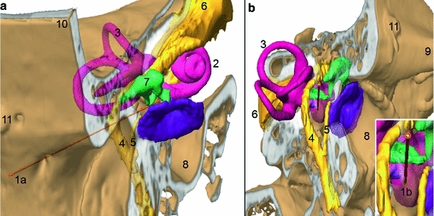
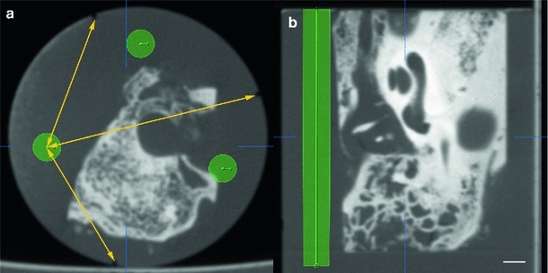
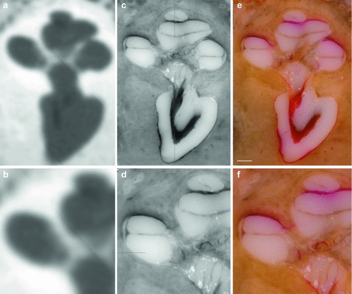
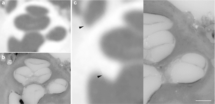
The image fusion of data from temporal bone specimens' and from non-destructive flat-panel-based volume computed tomography confirmed the spatial accuracy achieved by the procedure, as did the evaluation using the reference object.

CONCLUSION

This systematic and easy-to-follow preparation technique enables the three-dimensional (3D) histological reconstruction of complex soft and bony tissue structures. It facilitates the creation of detailed and spatially correct 3D anatomical models. Such models are of great benefit for image-based segmentation and planning in the field of computer-assisted surgery as well as in finite element analysis. In the context of human inner ear surgery, three-dimensional histology will improve the experimental evaluation and determination of intra-cochlear trauma after the insertion of an electrode array of a cochlear implant system.

摘要

目的

本文提出了一种高度精确的横断制备技术。研究目的是开发一种适用于具有高对比度和高分辨率的软、硬组织结构的成像方式。因此,追求了一种已经存在的微磨程序的改进。主要目标是保留空间关系,并确保对组织学切片进行准确的三维重建。

方法

使用了 12 个人类颞骨标本,包括中耳和内耳结构。它们被嵌入环氧树脂中,然后通过连续研磨进行解剖,最后进行数字化。使用具有 1 微米精度的触觉长度规测量每个研磨切片的实际磨损量。通过使用人工标记和应用基于特征的定制自动配准算法,将横截面图像对齐。为了确定整个重建过程的准确性,使用了一个已知的参考对象进行比较。为了确保组织学数据与常规临床图像数据的兼容性,最后将图像堆栈转换为 DICOM 标准。

结果

颞骨标本和无损平板式容积计算机断层扫描数据的图像融合证实了该程序达到的空间精度,使用参考对象的评估也是如此。

结论

这种系统且易于遵循的制备技术能够实现复杂的软、硬组织结构的三维(3D)组织学重建。它有助于创建详细且空间正确的 3D 解剖模型。在计算机辅助手术领域以及有限元分析中,这些模型对于基于图像的分割和规划非常有益。在人类内耳手术中,三维组织学将改善在插入人工耳蜗系统的电极阵列后对耳蜗内创伤的实验评估和确定。

https://cdn.ncbi.nlm.nih.gov/pmc/blobs/1dbf/3702969/3fdafe328580/11548_2013_825_Fig15_HTML.jpg
https://cdn.ncbi.nlm.nih.gov/pmc/blobs/1dbf/3702969/d8ca3e261d1e/11548_2013_825_Fig1_HTML.jpg
https://cdn.ncbi.nlm.nih.gov/pmc/blobs/1dbf/3702969/c8225a497242/11548_2013_825_Fig2_HTML.jpg
https://cdn.ncbi.nlm.nih.gov/pmc/blobs/1dbf/3702969/92ec687fb592/11548_2013_825_Fig3_HTML.jpg
https://cdn.ncbi.nlm.nih.gov/pmc/blobs/1dbf/3702969/544a3e16587f/11548_2013_825_Fig4_HTML.jpg
https://cdn.ncbi.nlm.nih.gov/pmc/blobs/1dbf/3702969/fb6401357fab/11548_2013_825_Fig5_HTML.jpg
https://cdn.ncbi.nlm.nih.gov/pmc/blobs/1dbf/3702969/47e913815360/11548_2013_825_Fig6_HTML.jpg
https://cdn.ncbi.nlm.nih.gov/pmc/blobs/1dbf/3702969/bc586fbe78ee/11548_2013_825_Fig7_HTML.jpg
https://cdn.ncbi.nlm.nih.gov/pmc/blobs/1dbf/3702969/6023e978ddb2/11548_2013_825_Fig8_HTML.jpg
https://cdn.ncbi.nlm.nih.gov/pmc/blobs/1dbf/3702969/a242e5dde97f/11548_2013_825_Fig9_HTML.jpg
https://cdn.ncbi.nlm.nih.gov/pmc/blobs/1dbf/3702969/433135f7cae8/11548_2013_825_Fig10_HTML.jpg
https://cdn.ncbi.nlm.nih.gov/pmc/blobs/1dbf/3702969/939200715205/11548_2013_825_Fig11_HTML.jpg
https://cdn.ncbi.nlm.nih.gov/pmc/blobs/1dbf/3702969/ab8a806ef9ae/11548_2013_825_Fig12_HTML.jpg
https://cdn.ncbi.nlm.nih.gov/pmc/blobs/1dbf/3702969/15d6bbb858f0/11548_2013_825_Fig13_HTML.jpg
https://cdn.ncbi.nlm.nih.gov/pmc/blobs/1dbf/3702969/62bc6192321b/11548_2013_825_Fig14_HTML.jpg
https://cdn.ncbi.nlm.nih.gov/pmc/blobs/1dbf/3702969/3fdafe328580/11548_2013_825_Fig15_HTML.jpg

相似文献

1
Three-dimensional histological specimen preparation for accurate imaging and spatial reconstruction of the middle and inner ear.用于中耳和内耳精确成像和空间重建的三维组织学标本制备。
Int J Comput Assist Radiol Surg. 2013 Jul;8(4):481-509. doi: 10.1007/s11548-013-0825-7. Epub 2013 Apr 30.
2
Characterizing the size of the target region for atraumatic opening of the cochlea through the facial recess.经面神经隐窝行耳蜗无创开窗时目标区域大小的特点。
Comput Med Imaging Graph. 2019 Oct;77:101655. doi: 10.1016/j.compmedimag.2019.101655. Epub 2019 Aug 30.
3
Diagnostic Value of 3D Segmentation in Understanding the Anatomy of Human Inner Ear Including Malformation Types.3D 分段对理解包括畸形类型在内的人类内耳解剖结构的诊断价值。
Ear Nose Throat J. 2021 Sep;100(5_suppl):675S-683S. doi: 10.1177/0145561320906621. Epub 2020 Feb 13.
4
Improved middle-ear soft-tissue visualization using synchrotron radiation phase-contrast imaging.使用同步辐射相衬成像改善中耳软组织可视化。
Hear Res. 2017 Oct;354:1-8. doi: 10.1016/j.heares.2017.08.001. Epub 2017 Aug 5.
5
Accuracy of computer-aided geometric 3D reconstruction based on histological serial microgrinding preparation.基于组织学连续微磨片制备的计算机辅助几何三维重建的准确性
Comput Methods Biomech Biomed Engin. 2011 Jul;14(7):581-94. doi: 10.1080/10255842.2010.487049.
6
Three-dimensional imaging of the temporal bone using a helical CT scan and its application in patients with cochlear implantation.使用螺旋CT扫描对颞骨进行三维成像及其在人工耳蜗植入患者中的应用。
ORL J Otorhinolaryngol Relat Spec. 1996 Nov-Dec;58(6):298-300. doi: 10.1159/000276857.
7
The creation of geometric three-dimensional models of the inner ear based on micro computer tomography data.基于微型计算机断层扫描数据创建内耳的几何三维模型。
Hear Res. 2008 Sep;243(1-2):95-104. doi: 10.1016/j.heares.2008.06.008. Epub 2008 Jun 28.
8
High-resolution computed tomography-based length assessments of the cochlea--an accuracy evaluation.基于高分辨率计算机断层扫描的耳蜗长度评估——准确性评价
Acta Otolaryngol. 2014 Oct;134(10):1011-5. doi: 10.3109/00016489.2014.913313.
9
Flat-panel volume computed tomography for cochlear implant electrode array examination in isolated temporal bone specimens.平板容积计算机断层扫描在孤立颞骨标本中用于人工耳蜗电极阵列检查
Otol Neurotol. 2006 Jun;27(4):491-8. doi: 10.1097/01.mao.0000194816.15298.50.
10
Evaluating Common Cavity Cochlear Deformities Using CT Images and 3D Reconstruction.使用 CT 图像和 3D 重建评估常见腔耳蜗畸形。
Laryngoscope. 2021 Feb;131(2):386-391. doi: 10.1002/lary.28640. Epub 2020 Apr 4.

引用本文的文献

1
Parameterisation and Prediction of Intra-canal Cochlear Structures.管内耳蜗结构的参数化与预测。
Ann Biomed Eng. 2024 Mar;52(3):695-706. doi: 10.1007/s10439-023-03417-5. Epub 2024 Jan 2.
2
Spatial correlation of 2D hard-tissue histology with 3D microCT scans through 3D printed phantoms.通过 3D 打印的体模实现二维硬组织组织学与三维 microCT 扫描的三维空间相关性。
Sci Rep. 2023 Oct 28;13(1):18479. doi: 10.1038/s41598-023-45518-0.
3
Current Approaches for Image Fusion of Histological Data with Computed Tomography and Magnetic Resonance Imaging.

本文引用的文献

1
Imaging procedures in cochlear implant patients - evaluation of different radiological techniques.人工耳蜗植入患者的成像程序——不同放射学技术的评估
Acta Otolaryngol. 2004 Apr;124 Suppl 552:46-9. doi: 10.1080/03655230410017175.
2
The OPFOS Microscopy Family: High-Resolution Optical Sectioning of Biomedical Specimens.OPFOS显微镜家族:生物医学标本的高分辨率光学切片
Anat Res Int. 2012;2012:206238. doi: 10.1155/2012/206238. Epub 2011 Nov 3.
3
Quantitative X-ray tomography of the mouse cochlea.小鼠耳蜗的定量 X 射线断层扫描。
将组织学数据与计算机断层扫描和磁共振成像进行图像融合的当前方法。
Radiol Res Pract. 2022 Nov 9;2022:6765895. doi: 10.1155/2022/6765895. eCollection 2022.
4
diceCT: A Valuable Technique to Study the Nervous System of Fish.体绘制 CT:研究鱼类神经系统的有效技术。
eNeuro. 2020 Aug 20;7(4). doi: 10.1523/ENEURO.0076-20.2020. Print 2020 Jul/Aug.
5
Visualizing the 3D cytoarchitecture of the human cochlea in an intact temporal bone using synchrotron radiation phase contrast imaging.利用同步辐射相衬成像技术在完整颞骨中可视化人类耳蜗的三维细胞结构。
Biomed Opt Express. 2018 Jul 18;9(8):3757-3767. doi: 10.1364/BOE.9.003757. eCollection 2018 Aug 1.
6
Enhanced contrast in X-ray microtomographic images of the membranous labyrinth using different X-ray sources and scanning modes.使用不同的 X 射线源和扫描模式增强膜迷路的 X 射线微断层图像的对比度。
J Anat. 2018 Dec;233(6):770-782. doi: 10.1111/joa.12885. Epub 2018 Oct 2.
7
Innovative 3D Model of the Human Middle Ear in High Resolution with a Histological Microgrinding Method: A Feasibility Study and Comparison with μCT.采用组织学微磨方法构建高分辨率人体中耳创新三维模型:一项可行性研究及与微计算机断层扫描的比较
Int J Otolaryngol. 2017;2017:6753604. doi: 10.1155/2017/6753604. Epub 2017 May 3.
8
Insertion forces and intracochlear trauma in temporal bone specimens implanted with a straight atraumatic electrode array.植入直形无创电极阵列的颞骨标本中的插入力和耳蜗内创伤
Eur Arch Otorhinolaryngol. 2017 May;274(5):2131-2140. doi: 10.1007/s00405-017-4485-z. Epub 2017 Feb 25.
9
Advanced Non-Destructive Ocular Visualization Methods by Improved X-Ray Imaging Techniques.通过改进的X射线成像技术实现的先进非破坏性眼部可视化方法。
PLoS One. 2017 Jan 27;12(1):e0170633. doi: 10.1371/journal.pone.0170633. eCollection 2017.
10
Visualization, measurement and modelling of the cochlea using rotating midmodiolar slice planes.使用旋转的中轴切片平面进行耳蜗的可视化、测量和建模。
Int J Comput Assist Radiol Surg. 2016 Oct;11(10):1855-69. doi: 10.1007/s11548-016-1374-7. Epub 2016 Mar 19.
PLoS One. 2012;7(4):e33568. doi: 10.1371/journal.pone.0033568. Epub 2012 Apr 2.
4
Three-dimensional representation of the human cochlea using micro-computed tomography data: presenting an anatomical model for further numerical calculations.利用微计算机断层扫描数据对人类耳蜗进行三维重建:提出一种用于进一步数值计算的解剖模型。
Acta Otolaryngol. 2012 Jun;132(6):603-13. doi: 10.3109/00016489.2011.653670. Epub 2012 Mar 4.
5
Open access high-resolution 3D morphology models of cat, gerbil, rabbit, rat and human ossicular chains.猫、沙鼠、兔、大鼠和人听小骨链的开放获取高分辨率 3D 形态模型。
Hear Res. 2012 Feb;284(1-2):1-5. doi: 10.1016/j.heares.2011.12.004. Epub 2011 Dec 13.
6
Optimising μCT imaging of the middle and inner cat ear.优化猫中耳和内耳的显微计算机断层扫描成像
Anat Histol Embryol. 2012 Apr;41(2):113-21. doi: 10.1111/j.1439-0264.2011.01110.x. Epub 2011 Oct 10.
7
Realistic 3D computer model of the gerbil middle ear, featuring accurate morphology of bone and soft tissue structures.沙鼠中耳的逼真 3D 计算机模型,具有精确的骨骼和软组织形态结构。
J Assoc Res Otolaryngol. 2011 Dec;12(6):681-96. doi: 10.1007/s10162-011-0281-4. Epub 2011 Jul 13.
8
Electric-acoustic stimulation of the auditory system: a review of the first decade.听觉系统的电声刺激:首个十年综述
Audiol Neurootol. 2011;16 Suppl 2:1-30. doi: 10.1159/000327765. Epub 2011 May 24.
9
TSLIM imaging and a morphometric analysis of the mouse spiral ganglion.TSLIM 成像及小鼠螺旋神经节的形态计量分析。
Hear Res. 2011 Aug;278(1-2):34-42. doi: 10.1016/j.heares.2011.02.008. Epub 2011 Mar 21.
10
Light sheet fluorescence microscopy: a review.光片荧光显微镜:综述。
J Histochem Cytochem. 2011 Feb;59(2):129-38. doi: 10.1369/0022155410394857.