Swarup J Shanti, Rao Arathi
Department of Paedodontics and Preventive Dentistry, Manipal College of Dental Sciences, Manipal University, Mangalore, India.
Contemp Clin Dent. 2012 Oct;3(4):433-6. doi: 10.4103/0976-237X.107434.
The purpose of this study was to evaluate the effects of synthetically processed hydroxyapatite particles in remineralization of the early enamel lesions in comparison with 2% sodium fluoride.
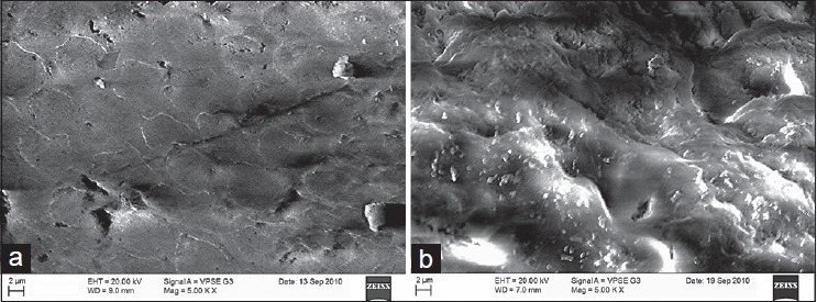
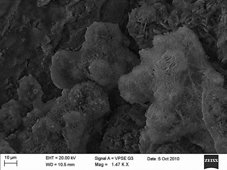
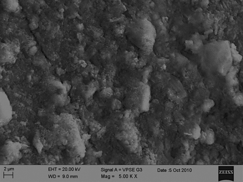
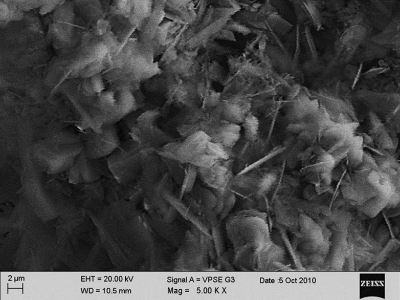
Thirty sound human premolars were divided into nanohydroxyapatite group (n = 15) and the sodium fluoride group (n = 15). The specimens were subjected to demineralization before being coated with 10% aqueous slurry of 20 nm nanohydroxyapatite or 2% sodium fluoride. The remineralizing efficacy of the materials was evaluated using surface microhardness (SMH) measurements, scanning microscopic analysis and analysis of the Ca/P ratio of the surface enamel. Data analysis was carried out using paired t-test and independent t-test.
The results showed that the nanohydroxyapatite group produced a surface morphology close to the biologic enamel, the increase in mineral content (Ca/P ratio) was more significant in the nanohydroxyapatite group (P < 0.05) and the SMH recovery was closer to the baseline level in the nanohydroxyapatite group (P < 0.05). Both the groups did not show any significant difference in thickness (P > 0.05).
The use of biomimetic nanohydroxyapatite as a remineralizing agent holds promise as a new synthetic enamel biocompatible material to repair early carious lesions.
本研究旨在评估合成处理的羟基磷灰石颗粒与2%氟化钠相比,对早期釉质病变再矿化的影响。
将30颗健康人前磨牙分为纳米羟基磷灰石组(n = 15)和氟化钠组(n = 15)。在涂覆20 nm纳米羟基磷灰石的10%水浆或2%氟化钠之前,先对标本进行脱矿处理。使用表面显微硬度(SMH)测量、扫描显微镜分析和表面釉质钙磷比分析来评估材料的再矿化效果。数据分析采用配对t检验和独立t检验。
结果表明,纳米羟基磷灰石组产生的表面形态接近生物釉质,纳米羟基磷灰石组的矿物质含量(钙磷比)增加更为显著(P < 0.05),且纳米羟基磷灰石组的SMH恢复更接近基线水平(P < 0.05)。两组在厚度方面均未显示出任何显著差异(P > 0.05)。
使用仿生纳米羟基磷灰石作为再矿化剂有望成为一种新型的合成釉质生物相容性材料,用于修复早期龋损。