Alant Jacob, Kemp Stephen, Webb Aubrey, Midha Rajiv
Hotchkiss Brain Institute and the Dept of Clinical Neurosciences, University of Calgary, Calgary, Alberta, Canada.
Evid Based Spine Care J. 2010 Aug;1(2):52-5. doi: 10.1055/s-0028-1100915.
Consistent with EBSJ's commitment to fostering quality research, we are pleased to feature some of the most highly rated abstracts from the 8th Annual AOSpine North America Fellows Forum in Banff Canada. Enhancing the quality of evidence in spine care means acknowledging and supporting the efforts of young researchers within our AOSpine North America network. We look forward to seeing more from these promising researchers in the future.
Basic science research report Introduction: Spinal nerve-injury management and prevention constitute a substantial proportion of a spinal surgeon's practice. Functional recovery after peripheral nerve injuries is often unsatisfactory and to optimize the outcomes, an intimate understanding of these injuries is required. Sunderland classified peripheral nerve injuries into five grades.1 Grade 1 (neurapraxia) and grade 2 (axonal disruption) injuries usually recover with no or insignificant functional deficits within weeks to a few months, respectively. Injuries that are most difficult to manage clinically are the often mixed grade 3 (endoneurial disruption) and grade 4 (perineurial disruption) lesions where spontaneous functional recovery is limited or absent, resulting in neuroma in continuity (NIC). Traumatic NIC is characterized by aberrant intra- and extra- fascicular axonal regeneration and scar formation within an unsevered injured nerve, resulting in impaired and erroneous end-organ reinnervation.2,3 Animal models reproducing grade 1, 2, 3, and 5 lesions have been developed, but to our knowledge a clinically relevant rodent model of NIC has not been developed.4,5,6,7,8 The effective peripheral nerve regeneration and resilience of rodents make it challenging to recreate the NIC scenario.
Our goal was to develop a practical rodent model for focal traumatic NIC, demonstrating the characteristic histological features, supported by concordant functional deficits. Such a model may help us to identify this injury pattern earlier and allow development of intervention strategies to reduce neuronal misdirection, scar formation, and enhance regeneration for improved functional recovery.
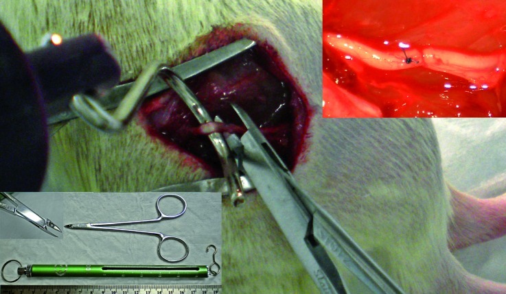
Various injury techniques were tested on freshly harvested Lewis rat sciatic nerves ex vivo, and examined histologically before inflicting more refined injuries in vivo. The optimal experimental injuries combined a 50 g traction force applied with a spring scale hooked around the sciatic nerve, and focal three second maximal compression using a malleus nipper (Figure 1). Nerves were harvested at 0, 5, 13, 21, and 65 days, and processed for longitudinal 8 micron cryostat sectioning, H&E, laminin, neurofilament, and Masson's trichrome staining. Skilled locomotion (tapered beam, ladder rung) and flat plane locomotion for ground reaction force (GRF) analysis were performed serially up to 9 weeks with the experimental (n = 4) and simple (control) crush (n = 1) injuries by blinded animal behavior experts, using methods as recently described.9 Figure 1 Photograph illustrating the experimental injury. Fifty grams of traction is applied in a direction orthogonal to the native nerve course after external neurolysis, simultaneously, three second maximal compression is applied at the sciatic trifurcation, just distal to a mesoneurial suture. Malleus nipper with tip detail and 100 g spring scale in bottom left. In situ sciatic nerve immediately after injury (top right).
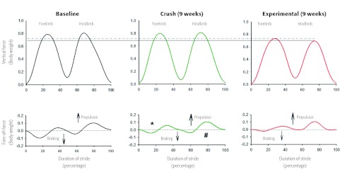
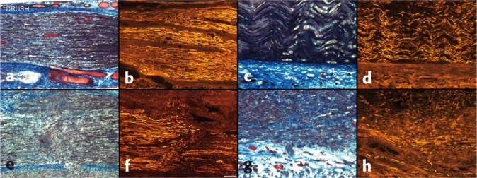
Disruption of the endoneurium and perineurium with aberrant intra- and extrafascicular axonal regeneration and progressive fibrosis was consistently demonstrated histologically in ten out of ten nerves with experimental injuries. In contrast, crush injuries showed only signs of Wallerian degeneration (Figure 2). At 8 weeks, experimental animals made more errors during skilled locomotion as compared to nerve crush animals. GRFs revealed impaired vertical and fore-aft force generation by the injured limbs at week 9 in the experimental group, whereas GRFs from the simple crush animal revealed recovery at the same time point (Figure 3). Figure 2 Injury zones at five days (a-d, bar = 200 µm) and 65 days (e-h, bar = 50 µm), comparing crush (top) to experimental (bottom) injuries; Masson's trichrome and neurofilament. Note the aberrant axonal sprouting and regeneration in the experimental injury group, associated with increased intrafascicular collagen, in contrast to orderly regeneration and lack of scar in the simple crush group.Figure 3 Mean vertical and fore-aft ground reaction forces at both baseline and 9 weeks from representative animals. Compared to baseline and crush-injured animal at 9 weeks, animals in the experimental group bear less weight on both their right (surgical) hind limb (solid line), and fore limb (dotted line) at 9 weeks. Comparable with historical data, the crush animal have improved braking ((*)) and propulsive (#) forces in fore and hind limbs (injured side) compared to the experimental group, though these have not returned to baseline values.
We have demonstrated histological features and poor functional recovery consistent with NIC formation in a rodent model. The injury mechanism employed combines traction and compression forces akin to the physical forces at play in clinical nerve injuries. Additional validating experiments are in progress.
与EBSJ致力于推动高质量研究的承诺相一致,我们很高兴展示第八届北美脊柱外科学会年会(于加拿大班夫举办)中一些评分最高的摘要。提高脊柱护理的证据质量意味着认可并支持北美脊柱外科学会网络内年轻研究人员的努力。我们期待未来能看到这些有前途的研究人员取得更多成果。
基础科学研究报告
脊髓神经损伤的管理与预防在脊柱外科医生的临床实践中占很大比例。周围神经损伤后的功能恢复往往不尽人意,为优化治疗效果,需要深入了解这些损伤。桑德兰将周围神经损伤分为五个等级。1级(神经失用症)和2级(轴突中断)损伤通常分别在数周内或数月内恢复,功能缺损不明显或无明显功能缺损。临床上最难处理的损伤通常是3级(神经内膜中断)和4级(神经束膜中断)的混合性损伤,自发功能恢复有限或不存在,导致连续性神经瘤(NIC)。创伤性NIC的特征是在未切断的损伤神经内,束内和束外轴突异常再生以及瘢痕形成,导致终末器官再支配受损和错误。2,3已经建立了可再现1、2、3和5级损伤的动物模型,但据我们所知,尚未开发出与临床相关的NIC啮齿动物模型。4,5,6,7,8啮齿动物有效的周围神经再生和恢复能力使得重现NIC情况具有挑战性。
我们的目标是开发一种实用的局灶性创伤性NIC啮齿动物模型,展示其特征性组织学特征,并伴有相应的功能缺损。这样的模型可能有助于我们更早地识别这种损伤模式,并开发干预策略以减少神经元错向、瘢痕形成,并促进再生以改善功能恢复。
在新鲜收获的Lewis大鼠坐骨神经上进行各种体外损伤技术测试,并在进行更精细的体内损伤之前进行组织学检查。最佳实验损伤包括用挂在坐骨神经上的弹簧秤施加50克牵引力,并使用锤状镊子进行三秒钟的局灶性最大压缩(图1)。在0、5、13、21和65天收获神经,并进行纵向8微米低温恒温器切片,并进行苏木精和伊红染色、层粘连蛋白染色、神经丝染色和Masson三色染色。由不知情的动物行为专家对实验性损伤组(n = 4)和简单(对照)挤压损伤组(n = 1)的动物连续进行长达9周的熟练运动(锥形梁、梯级)和平板运动以进行地面反作用力(GRF)分析,使用最近描述的方法。图1实验损伤示意图。在神经外膜松解后,在与神经自然走行方向正交的方向上施加50克牵引力,同时在坐骨神经分叉处(刚好在神经束膜缝合线的远端)进行三秒钟的最大压缩。左下角为带有尖端细节图的锤状镊子和100克弹簧秤。损伤后立即的坐骨神经原位图(右上角)。
在十根接受实验性损伤的神经中,有十根在组织学上始终显示神经内膜和神经束膜中断,伴有束内和束外轴突异常再生以及进行性纤维化。相比之下,挤压损伤仅显示沃勒变性的迹象(图2)。在第8周时,与神经挤压损伤的动物相比,实验动物在熟练运动中出现更多错误。GRF分析显示,在第9周时,实验组受伤肢体的垂直和前后力产生受损,而简单挤压损伤动物的GRF在同一时间点显示恢复(图3)。图2五天(a - d,标尺 = 200 µm)和65天(e - h,标尺 = 50 µm)时的损伤区域,比较挤压损伤(上图)和实验性损伤(下图);Masson三色染色和神经丝染色。注意实验性损伤组中异常的轴突发芽和再生,伴有束内胶原蛋白增加,而简单挤压组则是有序再生且无瘢痕形成。图3来自代表性动物在基线和9周时的平均垂直和前后地面反作用力。与基线和9周时挤压损伤的动物相比,实验组动物在第9周时右(手术)后肢(实线)和前肢(虚线)承受的重量均较轻。与历史数据相比,挤压损伤动物的前肢和后肢(受伤侧)的制动(*)和推进(#)力与实验组相比有所改善,尽管尚未恢复到基线值。
我们已经在啮齿动物模型中证明了与NIC形成一致的组织学特征和较差的功能恢复。所采用的损伤机制结合了牵引力和压缩力,类似于临床神经损伤中起作用的物理力。正在进行额外的验证实验。