Paterson Institute for Cancer Research, University of Manchester and Manchester Cancer Research Centre, Withington, Manchester, UK.
Cell Death Dis. 2013 May 2;4(5):e613. doi: 10.1038/cddis.2013.137.
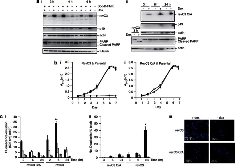
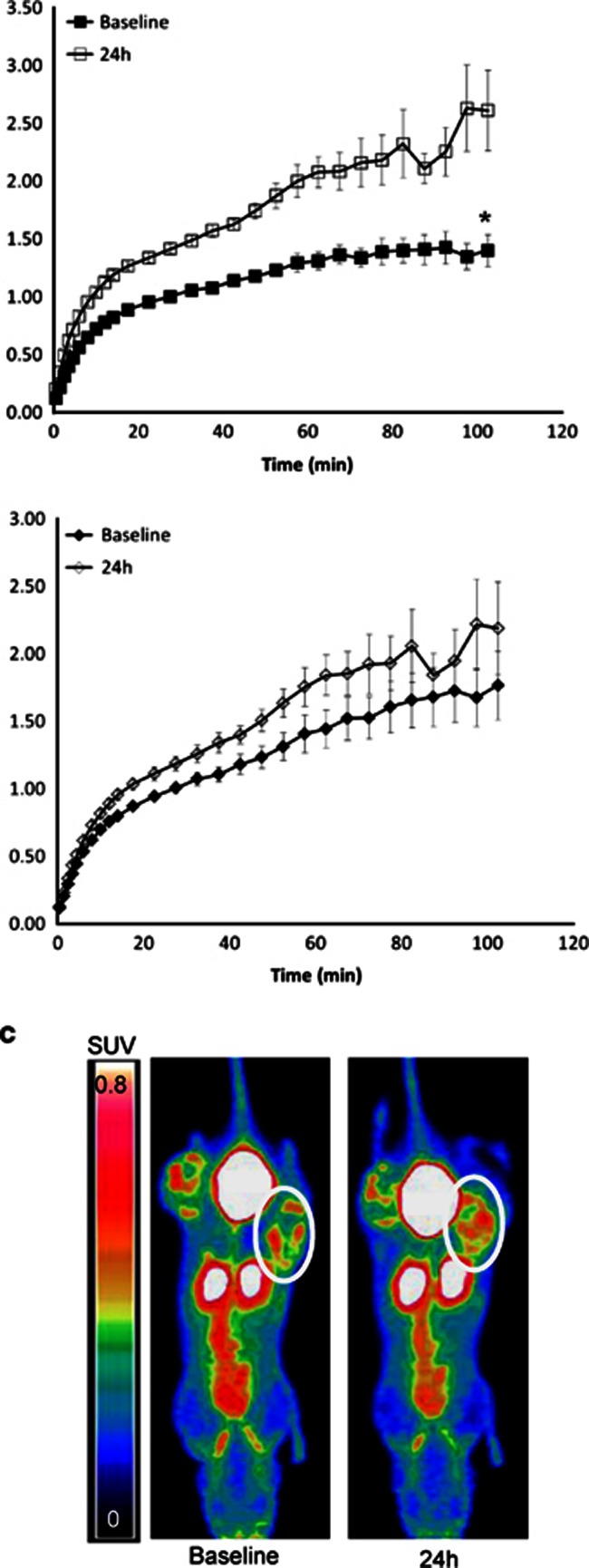
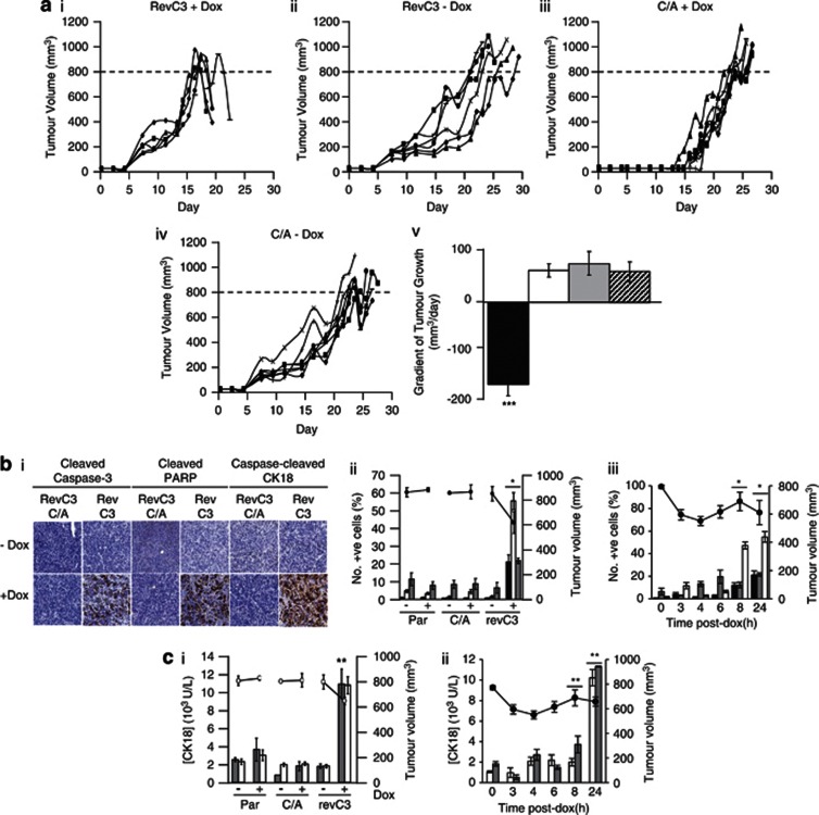
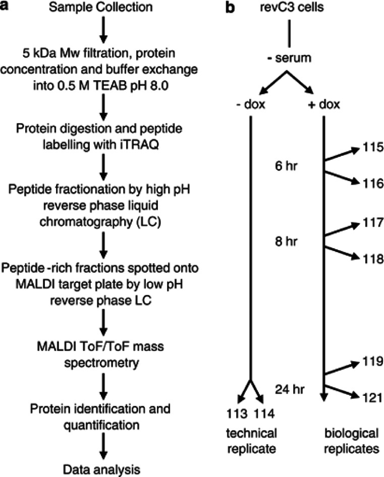
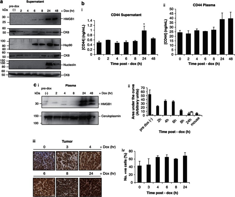
Novel anticancer drugs targeting key apoptosis regulators have been developed and are undergoing clinical trials. Pharmacodynamic biomarkers to define the optimum dose of drug that provokes tumor apoptosis are in demand; acquisition of longitudinal tumor biopsies is a significant challenge and minimally invasive biomarkers are required. Considering this, we have developed and validated a preclinical 'death-switch' model for the discovery of secreted biomarkers of tumour apoptosis using in vitro proteomics and in vivo evaluation of the novel imaging probe [(18)F]ML-10 for non-invasive detection of apoptosis using positron emission tomography (PET). The 'death-switch' is a constitutively active mutant caspase-3 that is robustly induced by doxycycline to drive synchronous apoptosis in human colorectal cancer cells in vitro or grown as tumor xenografts. Death-switch induction caused caspase-dependent apoptosis between 3 and 24 hours in vitro and regression of 'death-switched' xenografts occurred within 24 h correlating with the percentage of apoptotic cells in tumor and levels of an established cell death biomarker (cleaved cytokeratin-18) in the blood. We sought to define secreted biomarkers of tumor apoptosis from cultured cells using Discovery Isobaric Tag proteomics, which may provide candidates to validate in blood. Early after caspase-3 activation, levels of normally secreted proteins were decreased (e.g. Gelsolin and Midkine) and proteins including CD44 and High Mobility Group protein B1 (HMGB1) that were released into cell culture media in vitro were also identified in the bloodstream of mice bearing death-switched tumors. We also exemplify the utility of the death-switch model for the validation of apoptotic imaging probes using [(18)F]ML-10, a PET tracer currently in clinical trials. Results showed increased tracer uptake of [(18)F]ML-10 in tumours undergoing apoptosis, compared with matched tumour controls imaged in the same animal. Overall, the death-switch model represents a robust and versatile tool for the discovery and validation of apoptosis biomarkers.
针对关键凋亡调节剂的新型抗癌药物已经开发出来并正在进行临床试验。需要寻找药效生物标志物来确定引起肿瘤细胞凋亡的最佳药物剂量;获取纵向肿瘤活检是一个重大挑战,需要微创生物标志物。考虑到这一点,我们开发并验证了一种临床前的“死亡开关”模型,用于发现肿瘤细胞凋亡的分泌生物标志物,该模型使用体外蛋白质组学和新型成像探针 [(18)F]ML-10 在体内进行评估,用于使用正电子发射断层扫描 (PET) 进行非侵入性检测凋亡。“死亡开关”是一种组成型激活的突变胱天蛋白酶-3,它被强力霉素强烈诱导,以驱动体外人结直肠癌细胞或异种移植肿瘤中的同步凋亡。体外诱导死亡开关导致 caspase 依赖性凋亡发生在 3 至 24 小时之间,“死亡开关”异种移植物在 24 小时内消退,与肿瘤中凋亡细胞的百分比以及血液中已建立的细胞死亡生物标志物(裂解细胞角蛋白-18)的水平相关。我们试图使用 Discovery Isobaric Tag 蛋白质组学从培养细胞中定义肿瘤细胞凋亡的分泌生物标志物,这可能为验证血液中的生物标志物提供候选物。在 caspase-3 激活后不久,通常分泌的蛋白质水平降低(例如 Gelsolin 和 Midkine),并且在体外细胞培养物中释放到细胞培养基中的蛋白质,包括 CD44 和高迁移率族蛋白 B1(HMGB1),也在携带死亡开关肿瘤的小鼠血液中被鉴定出来。我们还通过使用目前正在临床试验中的 [(18)F]ML-10 验证凋亡成像探针,举例说明了死亡开关模型的实用性。结果显示,与在同一动物中成像的匹配肿瘤对照相比,正在凋亡的肿瘤中 [(18)F]ML-10 的摄取增加。总的来说,死亡开关模型代表了一种强大而通用的工具,可用于发现和验证凋亡生物标志物。