Medizinische Klinik und Poliklinik I, Klinikum der Universität München, Ludwig-Maximilians-Universität, Marchioninistrasse 15, 81377, Munich, Germany.
DZHK (German Centre for Cardiovascular Research), Partner Site Munich Heart Alliance, 80802, Munich, Germany.
Mol Imaging Biol. 2022 Aug;24(4):666-674. doi: 10.1007/s11307-022-01718-0. Epub 2022 Mar 29.
The loss of viable cardiac cells and cell death by myocardial infarction (MI) is still a significant obstacle in preventing deteriorating heart failure. Imaging of apoptosis, a defined cascade to cell death, could identify areas at risk.
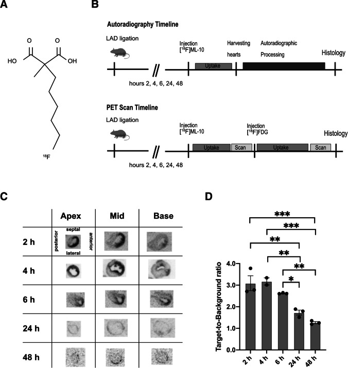
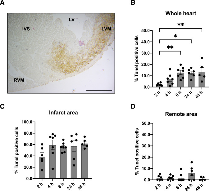
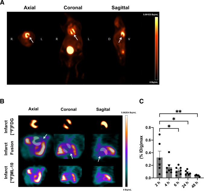
Using 2-(5-[F]fluoropentyl)-2-methyl-malonic acid ([F]ML-10) in autoradiography and positron emission tomography (PET) visualized apoptosis in murine hearts after permanent ligation of the left anterior descending artery (LAD) inducing myocardial infarction (MI). 2-deoxy-2-[F]fluoro-D-glucose ([F]FDG) PET imaging localized the infarct area after MI. Histology by terminal deoxynucleotidyl transferase dUTP nick end labeling (TUNEL) staining validated apoptosis in the heart.
Accumulation of [F]ML-10 was evident in the infarct area after permanent ligation of the LAD in autoradiography and PET imaging. Detection of apoptosis by [F]ML-10 is in line with the defect visualized by [F]FDG and the histological approach.
[F]ML-10 could be a suitable tracer for apoptosis imaging in a mouse model of permanent LAD ligation.
心肌梗死(MI)导致的存活心肌细胞丧失和细胞死亡仍然是预防心力衰竭恶化的重大障碍。细胞凋亡的成像,即细胞死亡的明确级联反应,可以识别出危险区域。
在永久性结扎左前降支(LAD)导致心肌梗死(MI)后,使用 2-(5-[F]氟戊基)-2-甲基丙二酸([F]ML-10)进行放射性自显影和正电子发射断层扫描(PET),在小鼠心脏中可视化细胞凋亡。2-脱氧-2-[F]氟-D-葡萄糖([F]FDG)PET 成像定位 MI 后的梗死区。末端脱氧核苷酸转移酶 dUTP 缺口末端标记(TUNEL)染色的组织学验证了心脏中的细胞凋亡。
在 LAD 永久性结扎后的放射性自显影和 PET 成像中,明显积聚了 [F]ML-10。[F]ML-10 检测到的细胞凋亡与 [F]FDG 和组织学方法可视化的缺陷一致。
[F]ML-10 可能是永久性 LAD 结扎的小鼠模型中细胞凋亡成像的合适示踪剂。