Department of Cardiology, Gelre Hospital, Albert Schweitzerlaan 31, 7334 DZ, Apeldoorn, the Netherlands,
Neth Heart J. 2013 Dec;21(12):561-4. doi: 10.1007/s12471-013-0426-7.
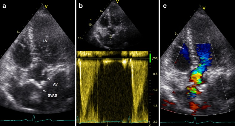
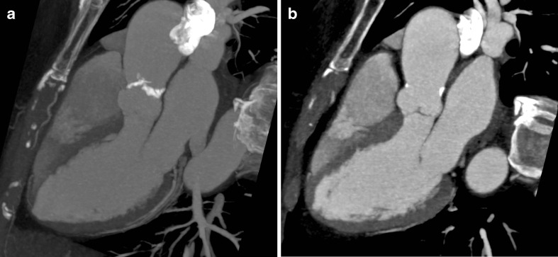
We report a case of a 64 year old woman with a calcified ring at the level of the sinotubular junction. Echocardiography and Computed Tomography showed a supravalvular aortic stenosis, without known associated lesions, except for the existence of an aberrant right subclavian artery. These combination of abnormalities makes it an unique case. Differential diagnosis of sinutubular calcification is added. From the literature a short review of supravalvular aortic stenosis is presented with indications for surgical intervention. Lifelong and regular follow up is necessary.
我们报告一例 64 岁女性患者,于窦管交界处存在环状钙化。超声心动图和计算机断层扫描显示瓣上型主动脉瓣狭窄,除存在右位锁骨下动脉异常外,无其他已知相关病变。这些异常的组合使其成为一个独特的病例。此外,还增加了窦管交界处钙化的鉴别诊断。从文献中简要回顾了瓣上型主动脉瓣狭窄,并提出了手术干预的适应证。需要终身和定期随访。