Department of Radiology, University of Washington Seattle, WA, USA ; Integrated Brain Imaging Center, University of Washington Seattle, WA, USA.
Front Neurol. 2013 May 1;4:43. doi: 10.3389/fneur.2013.00043. eCollection 2013.
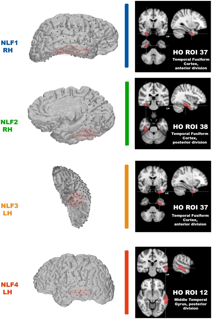
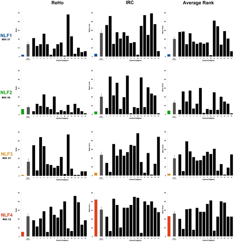
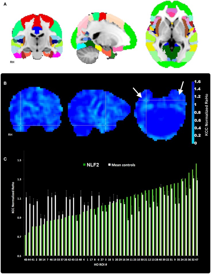
Successful resection of cortical tissue engendering seizure activity is efficacious for the treatment of refractory, focal epilepsy. The pre-operative localization of the seizure focus is therefore critical to yielding positive, post-operative outcomes. In a small proportion of focal epilepsy patients presenting with normal MRI, identification of the seizure focus is significantly more challenging. We examined the capacity of resting state functional MRI (rsfMRI) to identify the seizure focus in a group of four non-lesion, focal (NLF) epilepsy individuals. We predicted that computing patterns of local functional connectivity in and around the epileptogenic zone combined with a specific reference to the corresponding region within the contralateral hemisphere would reliably predict the location of the seizure focus. We first averaged voxel-wise regional homogeneity (ReHo) across regions of interest (ROIs) from a standardized, probabilistic atlas for each NLF subject as well as 16 age- and gender-matched controls. To examine contralateral effects, we computed a ratio of the mean pair-wise correlations of all voxels within a ROI with the corresponding contralateral region (IntraRegional Connectivity - IRC). For each subject, ROIs were ranked (from lowest to highest) on ReHo, IRC, and the mean of the two values. At the group level, we observed a significant decrease in the rank for ROI harboring the seizure focus for the ReHo rankings as well as for the mean rank. At the individual level, the seizure focus ReHo rank was within bottom 10% lowest ranked ROIs for all four NLF epilepsy patients and three out of the four for the IRC rankings. However, when the two ranks were combined (averaging across ReHo and IRC ranks and scalars), the seizure focus ROI was either the lowest or second lowest ranked ROI for three out of the four epilepsy subjects. This suggests that rsfMRI may serve as an adjunct pre-surgical tool, facilitating the identification of the seizure focus in focal epilepsy.
成功切除致痫性皮质组织对于治疗耐药性局灶性癫痫是有效的。因此,术前对致痫灶进行定位对于获得积极的术后效果至关重要。在一小部分表现为正常 MRI 的局灶性癫痫患者中,致痫灶的识别极具挑战性。我们检查了静息态功能磁共振成像(rsfMRI)在 4 名非病变性局灶性(NLF)癫痫患者中识别致痫灶的能力。我们预测,计算致痫区及其周围局部功能连接模式,并结合对侧半球相应区域的特定参考,将能够可靠地预测致痫灶的位置。我们首先对每个 NLF 患者以及 16 名年龄和性别匹配的对照者的标准化概率图谱的感兴趣区(ROI)进行了基于体素的区域同质性(ReHo)的平均计算。为了检查对侧效应,我们计算了 ROI 内所有体素的平均成对相关性与对应对侧区域的比值(区域内连接度 - IRC)。对于每个受试者,ROI 按 ReHo、IRC 和两者的平均值进行排序(从最低到最高)。在组水平上,我们观察到,对于 ReHo 评分和平均秩,包含致痫灶的 ROI 的秩显著降低。在个体水平上,四名 NLF 癫痫患者中,有四名患者的致痫灶 ReHo 评分排在所有 ROI 的倒数 10% 中,而 IRC 排名则为倒数 10%。然而,当将两个秩结合起来(对 ReHo 和 IRC 秩和标量进行平均)时,对于四个癫痫患者中的三个,致痫灶 ROI 要么是最低秩,要么是倒数第二低秩的 ROI。这表明 rsfMRI 可能成为一种辅助术前工具,有助于识别局灶性癫痫的致痫灶。