• 文献检索
  • 文档翻译
  • 深度研究
  • 学术资讯
  • Suppr Zotero 插件Zotero 插件
  • 邀请有礼
  • 套餐&价格
  • 历史记录
应用&插件
Suppr Zotero 插件Zotero 插件浏览器插件Mac 客户端Windows 客户端微信小程序
定价
高级版会员购买积分包购买API积分包
服务
文献检索文档翻译深度研究API 文档MCP 服务
关于我们
关于 Suppr公司介绍联系我们用户协议隐私条款
关注我们

Suppr 超能文献

核心技术专利:CN118964589B侵权必究
粤ICP备2023148730 号-1Suppr @ 2026

文献检索

告别复杂PubMed语法,用中文像聊天一样搜索,搜遍4000万医学文献。AI智能推荐,让科研检索更轻松。

立即免费搜索

文件翻译

保留排版,准确专业,支持PDF/Word/PPT等文件格式,支持 12+语言互译。

免费翻译文档

深度研究

AI帮你快速写综述,25分钟生成高质量综述,智能提取关键信息,辅助科研写作。

立即免费体验

Ki67、p16和CK17标记物在鉴别宫颈上皮内瘤变和良性病变中的评估

Evaluation of Ki67, p16 and CK17 Markers in Differentiating Cervical Intraepithelial Neoplasia and Benign Lesions.

作者信息

Sari Aslani Fatemeh, Safaei Akbar, Pourjabali Masoumeh, Momtahan Mozhdeh

机构信息

Department of Pathology, School of Medicine, Shiraz University of Medical Sciences, Shiraz, Iran;

出版信息

Iran J Med Sci. 2013 Mar;38(1):15-21.

PMID:23645953
原文链接:https://pmc.ncbi.nlm.nih.gov/articles/PMC3642940/
Abstract

BACKGROUND

Cervical intraepithelial neoplasia (CIN) is a premalignant lesion capable of progressing to cervical cancer. Despite the existing well-defined criteria, the histomorphologic diagnosis is subject to high rates of discordance among pathologists. The aim of this study was to evaluate Ki-67 (MIB-1), CK17 and p16 (INK4a) (p16) markers by immunohistochemical methods in differentiating CIN from benign cervical lesions.

METHODS

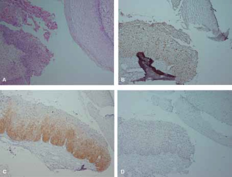
The present study reviewed and re-classified 77 cervical biopsies, originally diagnosed as 31 non-CIN, and 46 CIN, as 54 non-CIN, and 23 CIN based on at least two similar diagnoses. Immunostaining by Ki67, p16 and CK17 markers was performed on all cases and the results were compared with pervious and consensus diagnosis.

RESULTS

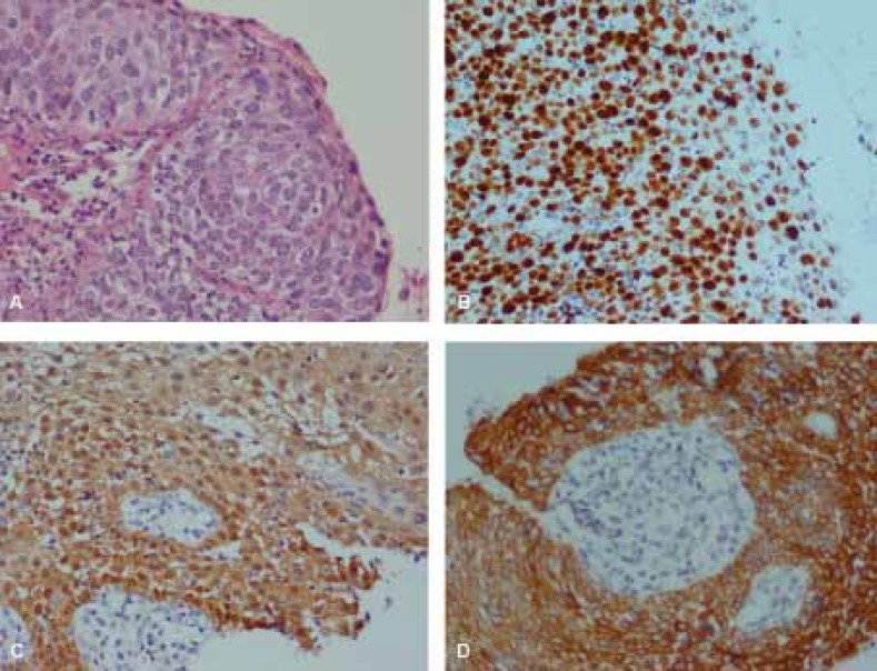
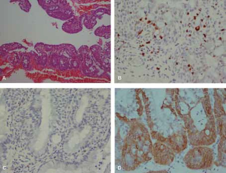
The overall agreement between pervious and consensus diagnosis was 67.5% (Kappa=0.39, P<0.001). The sensitivity and specificity of Ki67 immunostaining were 95.6% and 85.1% respectively, while for p16 the corresponding values were 91.3% and 98.1%. The overall agreement, for both p16 and Ki67, with consensus diagnosis were significant (P<0.001). The sensitivity and specificity of CK17 negative staining in CIN detection were 39.1% and 40.7% respectively.

CONCLUSION

Ki67 and p16 markers are recommended as complementary tests for differentiating between dysplastic and non-dysplastic lesions. CK17 does not discriminate between immature metaplasia with and without dysplasia.

摘要

背景

宫颈上皮内瘤变(CIN)是一种可发展为宫颈癌的癌前病变。尽管已有明确的诊断标准,但病理学家之间的组织形态学诊断存在较高的不一致率。本研究旨在通过免疫组化方法评估Ki-67(MIB-1)、CK17和p16(INK4a)(p16)标记物在鉴别CIN与良性宫颈病变中的作用。

方法

本研究对77例宫颈活检标本进行回顾性重新分类,最初诊断为31例非CIN和46例CIN,基于至少两种相似诊断重新分类为54例非CIN和23例CIN。对所有病例进行Ki67、p16和CK17标记物的免疫染色,并将结果与先前诊断和共识诊断进行比较。

结果

先前诊断与共识诊断之间的总体一致性为67.5%(Kappa=0.39,P<0.001)。Ki67免疫染色的敏感性和特异性分别为95.6%和85.1%,而p16的相应值分别为91.3%和98.1%。p16和Ki67与共识诊断的总体一致性均具有显著性(P<0.001)。CK17阴性染色在CIN检测中的敏感性和特异性分别为39.1%和40.7%。

结论

推荐将Ki67和p16标记物作为鉴别发育异常和非发育异常病变的补充检查。CK17不能区分有无发育异常的未成熟化生。

https://cdn.ncbi.nlm.nih.gov/pmc/blobs/ca11/3642940/10ac179de96a/IJMS-038-015-g003.jpg
https://cdn.ncbi.nlm.nih.gov/pmc/blobs/ca11/3642940/e6b096b220a4/IJMS-038-015-g001.jpg
https://cdn.ncbi.nlm.nih.gov/pmc/blobs/ca11/3642940/ca0254f1e123/IJMS-038-015-g002.jpg
https://cdn.ncbi.nlm.nih.gov/pmc/blobs/ca11/3642940/10ac179de96a/IJMS-038-015-g003.jpg
https://cdn.ncbi.nlm.nih.gov/pmc/blobs/ca11/3642940/e6b096b220a4/IJMS-038-015-g001.jpg
https://cdn.ncbi.nlm.nih.gov/pmc/blobs/ca11/3642940/ca0254f1e123/IJMS-038-015-g002.jpg
https://cdn.ncbi.nlm.nih.gov/pmc/blobs/ca11/3642940/10ac179de96a/IJMS-038-015-g003.jpg

相似文献

1
Evaluation of Ki67, p16 and CK17 Markers in Differentiating Cervical Intraepithelial Neoplasia and Benign Lesions.Ki67、p16和CK17标记物在鉴别宫颈上皮内瘤变和良性病变中的评估
Iran J Med Sci. 2013 Mar;38(1):15-21.
2
Immunohistochemical Study of p16INK4A, MIB-1 and CK17 in Pre-neoplastic and Neoplastic Epithelial Lesions of Cervix.p16INK4A、MIB-1 和 CK17 在宫颈上皮内瘤变和肿瘤性病变中的免疫组化研究。
Gulf J Oncolog. 2022 Sep;1(40):29-37.
3
CK17 and p16 expression patterns distinguish (atypical) immature squamous metaplasia from high-grade cervical intraepithelial neoplasia (CIN III).细胞角蛋白17(CK17)和p16的表达模式可将(非典型)不成熟鳞状化生与高级别宫颈上皮内瘤变(CIN III)区分开来。
Histopathology. 2007 Apr;50(5):629-35. doi: 10.1111/j.1365-2559.2007.02652.x.
4
Role of p16, CK17, p63, and human papillomavirus in diagnosis of cervical intraepithelial neoplasia and distinction from its mimics.p16、细胞角蛋白17(CK17)、p63及人乳头瘤病毒在宫颈上皮内瘤变诊断及其与相似病变鉴别中的作用
Int J Surg Pathol. 2014 May;22(3):221-30. doi: 10.1177/1066896913496147. Epub 2013 Jul 24.
5
Coordinate expression of cytokeratin 8 and cytokeratin 17 immunohistochemical staining in cervical intraepithelial neoplasia and cervical squamous cell carcinoma: an immunohistochemical analysis and review of the literature.细胞角蛋白8和细胞角蛋白17免疫组化染色在宫颈上皮内瘤变和宫颈鳞状细胞癌中的协同表达:一项免疫组化分析及文献综述
Gynecol Oncol. 2008 Mar;108(3):598-602. doi: 10.1016/j.ygyno.2007.11.042. Epub 2008 Jan 14.
6
Expression of P63, P16 and CK17 in Atypical Squamous Metaplasia and Cervical Intraepithelial Neoplasia.P63、P16和CK17在非典型鳞状化生及宫颈上皮内瘤变中的表达
Iran J Pathol. 2021 Spring;16(2):181-189. doi: 10.30699/IJP.2021.104280.2095. Epub 2020 Dec 26.
7
Role of p16/INK4a and Ki-67 as specific biomarkers for cervical intraepithelial neoplasia: An institutional study.p16/INK4a和Ki-67作为宫颈上皮内瘤变特异性生物标志物的作用:一项机构研究。
J Lab Physicians. 2017 Apr-Jun;9(2):104-110. doi: 10.4103/0974-2727.199630.
8
Correlation of P16 (Ink4a) and CK17 to HPV (16E6+18E6) in Premalignant and Malignant Lesions of Uterine Cervix: A Clinicopathologic Study.子宫颈癌前病变和恶性病变中P16(Ink4a)及细胞角蛋白17与HPV(16E6 + 18E6)的相关性:一项临床病理研究
Iran J Pathol. 2016 Fall;11(4):377-390.
9
Re-evaluation of Negative Cone Biopsy Results with Ki-67 and p16 Immunostaining following Positive Cervical Biopsy.宫颈活检阳性后,用Ki-67和p16免疫染色对阴性锥形活检结果进行重新评估。
Iran J Med Sci. 2020 Nov;45(6):469-476. doi: 10.30476/ijms.2020.72707.0.
10
Comparison of Ki67 index and P16 expression in different grades of cervical squamous intraepithelial lesions.不同级别宫颈鳞状上皮内病变中Ki67指数与P16表达的比较。
Caspian J Intern Med. 2023 Winter;14(1):69-75. doi: 10.22088/cjim.14.1.69.

引用本文的文献

1
Comparison of Ki67 index and P16 expression in different grades of cervical squamous intraepithelial lesions.不同级别宫颈鳞状上皮内病变中Ki67指数与P16表达的比较。
Caspian J Intern Med. 2023 Winter;14(1):69-75. doi: 10.22088/cjim.14.1.69.
2
The value of Ki67 for the diagnosis of LSIL and the problems of p16 in the diagnosis of HSIL.Ki67 在 LSIL 诊断中的价值及 p16 在 HSIL 诊断中的问题。
Sci Rep. 2022 May 9;12(1):7613. doi: 10.1038/s41598-022-11584-z.
3
HIV-Associated Cancer Biomarkers: A Requirement for Early Diagnosis.

本文引用的文献

1
Evaluation of cervical cone biopsies for coexpression of p16INK4a and Ki-67 in epithelial cells.评估宫颈锥切标本中上皮细胞 p16INK4a 和 Ki-67 的共表达。
Int J Cancer. 2012 Jan 15;130(2):388-94. doi: 10.1002/ijc.26017. Epub 2011 Jun 10.
2
p16(INK4a) immunostaining as an alternative to histology review for reliable grading of cervical intraepithelial lesions.p16(INK4a)免疫染色作为替代组织学复查用于可靠分级宫颈上皮内病变。
J Clin Pathol. 2010 Nov;63(11):972-7. doi: 10.1136/jcp.2010.078634. Epub 2010 Oct 5.
3
Using biomarkers as objective standards in the diagnosis of cervical biopsies.
HIV 相关癌症生物标志物:早期诊断的需要。
Int J Mol Sci. 2021 Jul 29;22(15):8127. doi: 10.3390/ijms22158127.
4
Predictive Value of Marker of Proliferation Ki-67 and Cell Cycle Dependent Protein kinase Inhibitor P16INK4a in Cervical Biopsy to Determine Its Biological Behaviour.Ki-67 增殖标志物和细胞周期依赖性蛋白激酶抑制剂 P16INK4a 在宫颈活检中预测其生物学行为的价值。
Asian Pac J Cancer Prev. 2021 Jul 1;22(7):2237-2241. doi: 10.31557/APJCP.2021.22.7.2237.
5
Expression of P63, P16 and CK17 in Atypical Squamous Metaplasia and Cervical Intraepithelial Neoplasia.P63、P16和CK17在非典型鳞状化生及宫颈上皮内瘤变中的表达
Iran J Pathol. 2021 Spring;16(2):181-189. doi: 10.30699/IJP.2021.104280.2095. Epub 2020 Dec 26.
6
Evaluation of Immunohistochemical Markers, CK17 and SOX2, as Adjuncts to p53 for the Diagnosis of Differentiated Vulvar Intraepithelial Neoplasia (dVIN).评估免疫组织化学标志物CK17和SOX2作为p53的辅助指标用于诊断分化型外阴上皮内瘤变(dVIN)。
Pharmaceuticals (Basel). 2021 Apr 2;14(4):324. doi: 10.3390/ph14040324.
7
Relationship of P16 and Ki67 in recurrence of HPV infection and cervical intraepithelial neoplasia.P16与Ki67在人乳头瘤病毒感染复发及宫颈上皮内瘤变中的关系
Int J Clin Exp Pathol. 2020 Dec 1;13(12):3174-3180. eCollection 2020.
8
Evaluation of the Performance of CinTec® PLUS in SurePathTM Liquid-Based Cervico-Vaginal Samples.评估 CinTec® PLUS 在 SurePathTM 液基宫颈阴道样本中的性能。
Turk Patoloji Derg. 2021;37(1):32-38. doi: 10.5146/tjpath.2020.01505.
9
Re-evaluation of Negative Cone Biopsy Results with Ki-67 and p16 Immunostaining following Positive Cervical Biopsy.宫颈活检阳性后,用Ki-67和p16免疫染色对阴性锥形活检结果进行重新评估。
Iran J Med Sci. 2020 Nov;45(6):469-476. doi: 10.30476/ijms.2020.72707.0.
10
Bioinformatics analysis and identification of potential genes related to pathogenesis of cervical intraepithelial neoplasia.宫颈上皮内瘤变发病机制相关潜在基因的生物信息学分析与鉴定
J Cancer. 2020 Feb 3;11(8):2150-2157. doi: 10.7150/jca.38211. eCollection 2020.
在宫颈活检诊断中使用生物标志物作为客观标准。
Am J Surg Pathol. 2010 Aug;34(8):1077-87. doi: 10.1097/PAS.0b013e3181e8b2c4.
4
Conjunctive p16INK4a testing significantly increases accuracy in diagnosing high-grade cervical intraepithelial neoplasia.联合检测 p16INK4a 可显著提高诊断高级别宫颈上皮内瘤变的准确性。
Am J Clin Pathol. 2010 Mar;133(3):395-406. doi: 10.1309/AJCPXSVCDZ3D5MZM.
5
Value of p16(INK4a) as a marker of progression/regression in cervical intraepithelial neoplasia grade 1.p16(INK4a)作为1级宫颈上皮内瘤变进展/消退标志物的价值
Am J Obstet Gynecol. 2009 Nov;201(5):488.e1-7. doi: 10.1016/j.ajog.2009.05.046. Epub 2009 Aug 15.
6
Expression of the p16 and Ki-67 in relation to the grade of cervical intraepithelial neoplasia and high-risk human papillomavirus infection.p16 和 Ki-67 的表达与宫颈上皮内瘤变分级和高危型人乳头瘤病毒感染的关系。
J Gynecol Oncol. 2008 Sep;19(3):162-8. doi: 10.3802/jgo.2008.19.3.162. Epub 2008 Sep 30.
7
Human papillomavirus mRNA and p16 detection as biomarkers for the improved diagnosis of cervical neoplasia.人乳头瘤病毒mRNA和p16检测作为改善宫颈肿瘤诊断的生物标志物。
Cancer Epidemiol Biomarkers Prev. 2008 Oct;17(10):2536-45. doi: 10.1158/1055-9965.EPI-08-0306.
8
Immunostaining for p16INK4a used as a conjunctive tool improves interobserver agreement of the histologic diagnosis of cervical intraepithelial neoplasia.将p16INK4a免疫染色用作联合工具可提高宫颈上皮内瘤变组织学诊断的观察者间一致性。
Am J Surg Pathol. 2008 Apr;32(4):502-12. doi: 10.1097/PAS.0b013e31815ac420.
9
Coordinate expression of cytokeratin 8 and cytokeratin 17 immunohistochemical staining in cervical intraepithelial neoplasia and cervical squamous cell carcinoma: an immunohistochemical analysis and review of the literature.细胞角蛋白8和细胞角蛋白17免疫组化染色在宫颈上皮内瘤变和宫颈鳞状细胞癌中的协同表达:一项免疫组化分析及文献综述
Gynecol Oncol. 2008 Mar;108(3):598-602. doi: 10.1016/j.ygyno.2007.11.042. Epub 2008 Jan 14.
10
p16 and Ki-67 immunostaining in atypical immature squamous metaplasia of the uterine cervix: correlation with human papillomavirus detection.p16和Ki-67免疫染色在子宫颈非典型未成熟鳞状化生中的应用:与人乳头瘤病毒检测的相关性
Arch Pathol Lab Med. 2007 Sep;131(9):1343-9. doi: 10.5858/2007-131-1343-PAKIIA.