School of Nano-Bioscience and Chemical Engineering, Ulsan National Institute of Science and Technology (UNIST), Ulsan 689-798, Korea.
J Neural Eng. 2013 Jun;10(3):036020. doi: 10.1088/1741-2560/10/3/036020. Epub 2013 May 8.
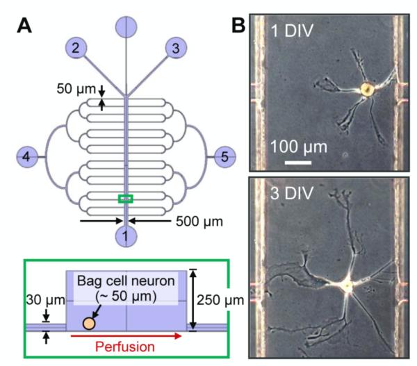
The regeneration and repair of damaged neuronal networks is a difficult process to study in vivo, leading to the development of multiple in vitro models and techniques for studying nerve injury. Here we describe an approach for generating a well-defined subcellular neurite injury in a microfluidic device.
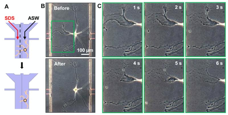
A defined laminar stream of sodium dodecyl sulfate (SDS) was used to damage selected portions of neurites of individual neurons. The somata and neurites unaffected by the SDS stream remained viable, thereby enabling the study of neuronal regeneration.
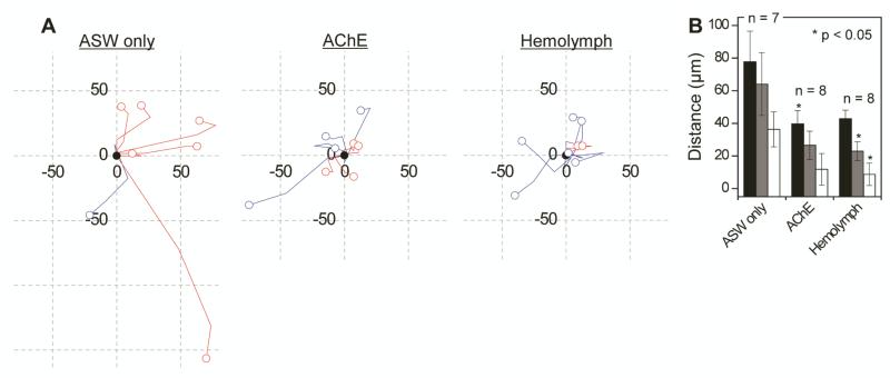
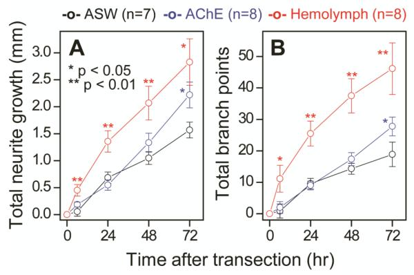
By using well-characterized neurons from Aplysia californica cultured in vitro, we demonstrate that our approach is useful in creating neurite damage, investigating neurotrophic factors, and monitoring somata migration during regeneration. Supplementing the culture medium with acetylcholinesterase (AChE) or Aplysia hemolymph facilitated the regeneration of the peptidergic Aplysia neurons within 72 h, with longer (p < 0.05) and more branched (p < 0.05) neurites than in the control medium. After the neurons were transected, their somata migrated; intriguingly, for the control cultures, the migration direction was always away from the injury site (7/7). In the supplemented cultures, the number decreased to 6/8 in AChE and 4/8 in hemolymph, with reduced migration distances in both cases.
The SDS transection approach is simple and inexpensive, yet provides flexibility in studying neuroregeneration, particularly when it is important to make sure there are no retrograde signals from the distal segments affecting regeneration. Neurons are known to not only be under tension but also balanced in terms of force, and the balance is obviously disrupted by transection. Our experimental platform, verified with Aplysia, can be extended to mammalian systems, and help us gain insight into the role that neurotrophic factors and mechanical tension play during neuronal regeneration.
在体内研究受损神经元网络的再生和修复是一个困难的过程,导致了多种体外模型和技术的发展,用于研究神经损伤。在这里,我们描述了一种在微流控装置中产生明确定义的亚细胞轴突损伤的方法。
使用十二烷基硫酸钠(SDS)的定义层流来损伤单个神经元的选定部分轴突。未受 SDS 流影响的神经元体和轴突保持存活,从而能够研究神经元再生。
通过使用体外培养的加利福尼亚海兔(Aplysia californica)的特征明确的神经元,我们证明我们的方法可用于创建轴突损伤,研究神经营养因子,并在再生过程中监测神经元体迁移。在培养基中补充乙酰胆碱酯酶(AChE)或海兔血淋巴促进了肽能 Aplysia 神经元在 72 小时内的再生,其轴突比对照培养基中更长(p<0.05)且更分支(p<0.05)。神经元被切断后,其神经元体迁移;有趣的是,对于对照培养物,迁移方向总是远离损伤部位(7/7)。在补充培养物中,AChE 中的数量减少到 6/8,在血淋巴中减少到 4/8,两种情况下的迁移距离都减少了。
SDS 横切方法简单且廉价,但在研究神经再生方面具有灵活性,特别是在确保没有来自远端段的逆行信号影响再生时。众所周知,神经元不仅受到张力的影响,而且在力方面也保持平衡,而这种平衡显然会被横切破坏。我们的实验平台,经过 Aplysia 验证,可以扩展到哺乳动物系统,并帮助我们深入了解神经营养因子和机械张力在神经元再生过程中所起的作用。