National Research Council, Fort Sam Houston, Texas, USA.
PLoS One. 2013 Apr 29;8(4):e63122. doi: 10.1371/journal.pone.0063122. Print 2013.
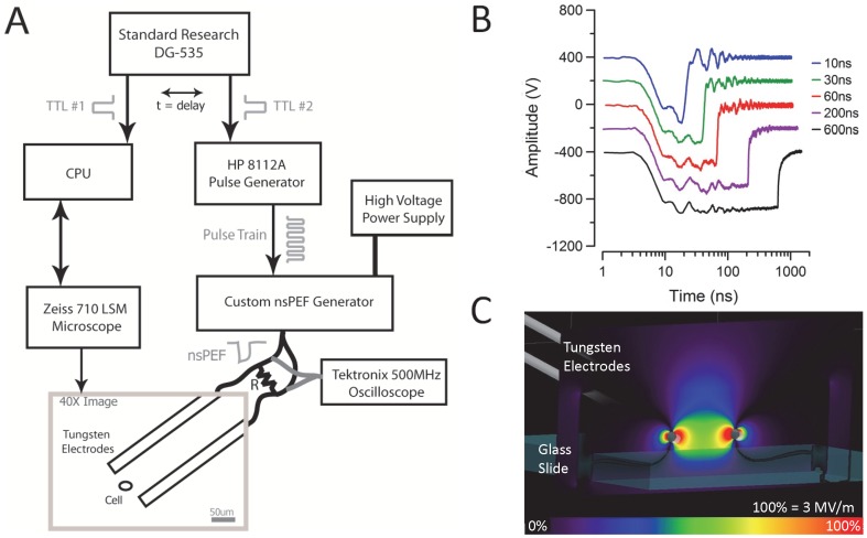
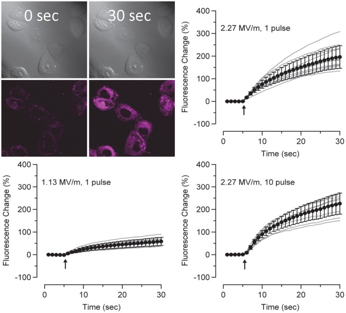
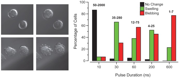
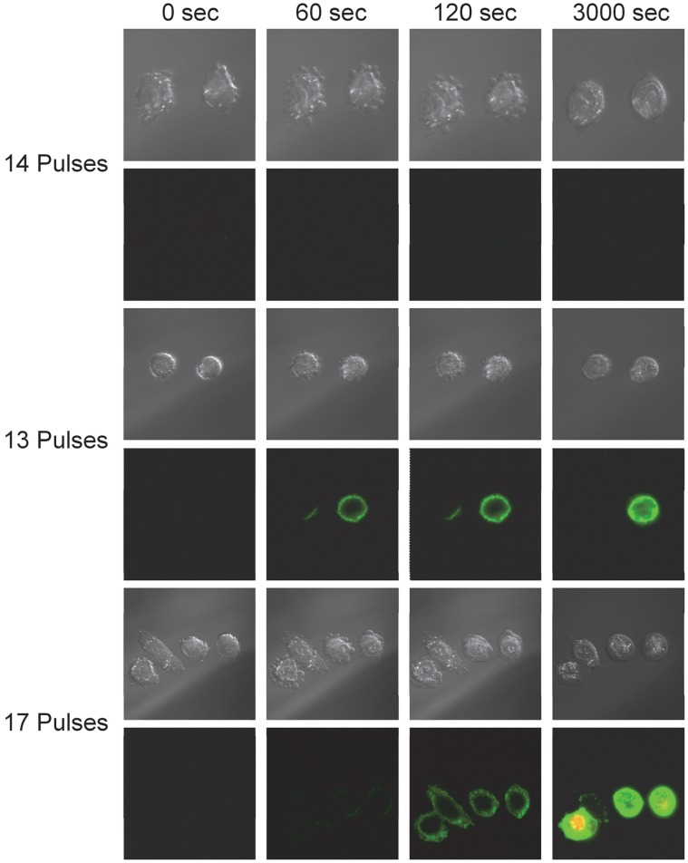
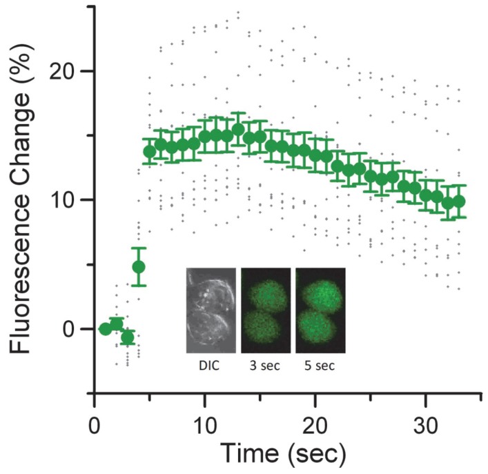
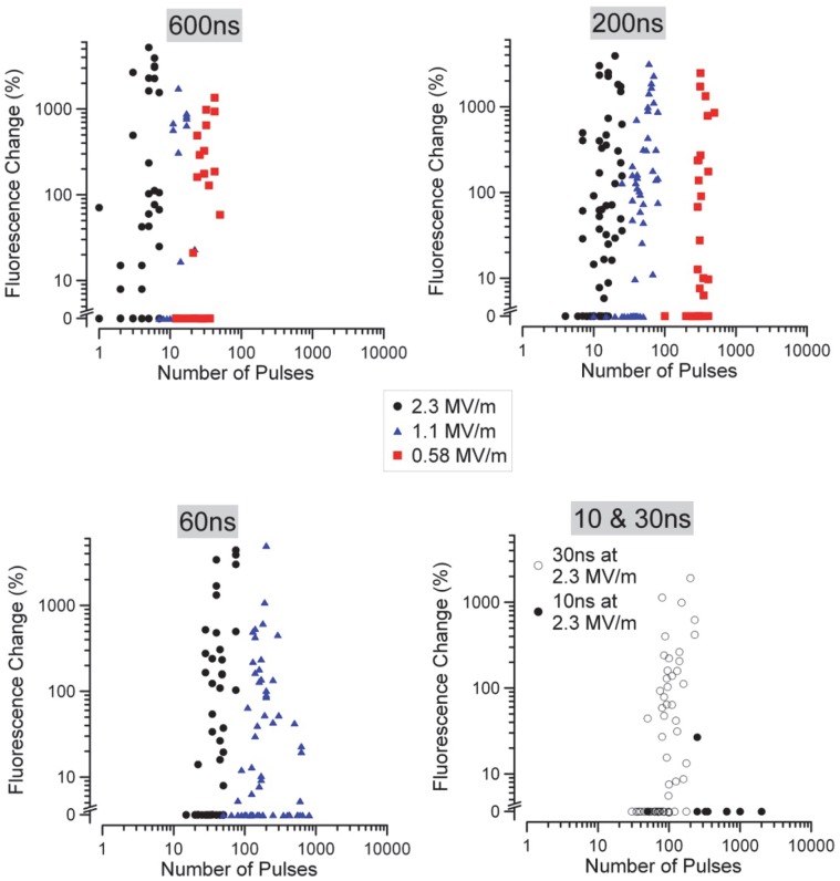
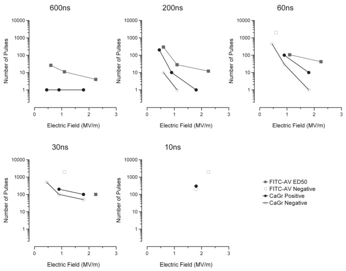
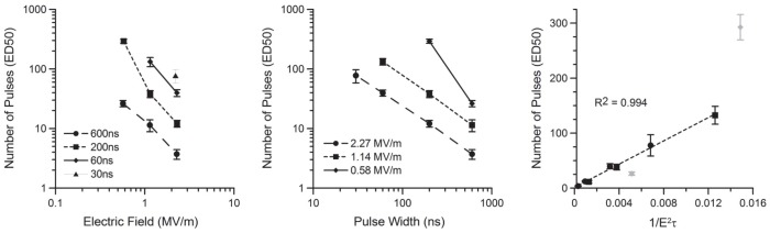
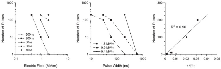
High-amplitude, MV/m, nanosecond pulsed electric fields (nsPEF) have been hypothesized to cause nanoporation of the plasma membrane. Phosphatidylserine (PS) externalization has been observed on the outer leaflet of the membrane shortly after nsPEF exposure, suggesting local structural changes in the membrane. In this study, we utilized fluorescently-tagged Annexin V to observe the externalization of PS on the plasma membrane of isolated Chinese Hamster Ovary (CHO) cells following exposure to nsPEF. A series of experiments were performed to determine the dosimetric trends of PS expression caused by nsPEF as a function of pulse duration, τ, delivered field strength, ED, and pulse number, n. To accurately estimate dose thresholds for cellular response, data were reduced to a set of binary responses and ED50s were estimated using Probit analysis. Probit analysis results revealed that PS externalization followed the non-linear trend of (τ*ED (2))(-1) for high amplitudes, but failed to predict low amplitude responses. A second set of experiments was performed to determine the nsPEF parameters necessary to cause observable calcium uptake, using cells preloaded with calcium green (CaGr), and membrane permeability, using FM1-43 dye. Calcium influx and FM1-43 uptake were found to always be observed at lower nsPEF exposure parameters compared to PS externalization. These findings suggest that multiple, higher amplitude and longer pulse exposures may generate pores of larger diameter enabling lateral diffusion of PS; whereas, smaller pores induced by fewer, lower amplitude and short pulse width exposures may only allow extracellular calcium and FM1-43 uptake.
高振幅、MV/m、纳秒级脉冲电场(nsPEF)被假设会导致质膜纳米孔化。在 nsPEF 暴露后不久,就观察到磷脂酰丝氨酸(PS)在膜的外叶外侧化,这表明膜的局部结构发生了变化。在这项研究中,我们利用荧光标记的 Annexin V 观察到 nsPEF 暴露后分离的中国仓鼠卵巢(CHO)细胞质膜上 PS 的外溢。进行了一系列实验,以确定 nsPEF 引起的 PS 表达的剂量趋势,作为脉冲持续时间 τ、施加的场强 ED 和脉冲数 n 的函数。为了准确估计细胞反应的剂量阈值,将数据简化为一组二进制响应,并使用 Probit 分析估计 ED50。Probit 分析结果表明,PS 外溢遵循(τ*ED(2))(-1)的非线性趋势,对于高振幅,但未能预测低振幅响应。进行了第二组实验,以确定引起可观察到的钙摄取所需的 nsPEF 参数,使用预先加载钙绿(CaGr)的细胞,并使用 FM1-43 染料确定膜通透性。发现钙内流和 FM1-43 摄取始终在低于 PS 外溢的 nsPEF 暴露参数下观察到。这些发现表明,多个更高幅度和更长脉冲的暴露可能会产生更大直径的孔,从而允许 PS 侧向扩散;而较少、幅度较低和短脉冲宽度的暴露产生的较小孔可能只允许细胞外钙和 FM1-43 摄取。