• 文献检索
  • 文档翻译
  • 深度研究
  • 学术资讯
  • Suppr Zotero 插件Zotero 插件
  • 邀请有礼
  • 套餐&价格
  • 历史记录
应用&插件
Suppr Zotero 插件Zotero 插件浏览器插件Mac 客户端Windows 客户端微信小程序
定价
高级版会员购买积分包购买API积分包
服务
文献检索文档翻译深度研究API 文档MCP 服务
关于我们
关于 Suppr公司介绍联系我们用户协议隐私条款
关注我们

Suppr 超能文献

核心技术专利:CN118964589B侵权必究
粤ICP备2023148730 号-1Suppr @ 2026

文献检索

告别复杂PubMed语法,用中文像聊天一样搜索,搜遍4000万医学文献。AI智能推荐,让科研检索更轻松。

立即免费搜索

文件翻译

保留排版,准确专业,支持PDF/Word/PPT等文件格式,支持 12+语言互译。

免费翻译文档

深度研究

AI帮你快速写综述,25分钟生成高质量综述,智能提取关键信息,辅助科研写作。

立即免费体验

标记融合:细胞-细胞融合中的磷脂酰丝氨酸信号。

Flagging fusion: Phosphatidylserine signaling in cell-cell fusion.

机构信息

Section on Membrane Biology, Eunice Kennedy Shriver National Institute of Child Health and Human Development, National Institutes of Health, Bethesda, Maryland, USA.

Section on Membrane Biology, Eunice Kennedy Shriver National Institute of Child Health and Human Development, National Institutes of Health, Bethesda, Maryland, USA.

出版信息

J Biol Chem. 2021 Jan-Jun;296:100411. doi: 10.1016/j.jbc.2021.100411. Epub 2021 Feb 11.

DOI:10.1016/j.jbc.2021.100411
PMID:33581114
原文链接:https://pmc.ncbi.nlm.nih.gov/articles/PMC8005811/
Abstract

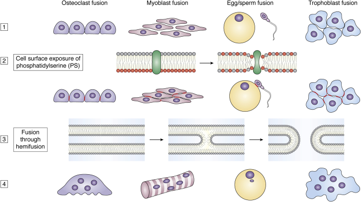
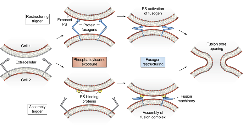
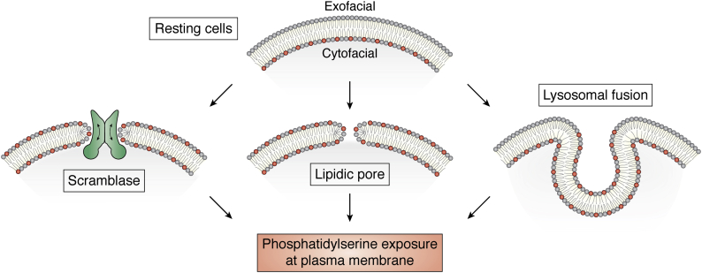
Formations of myofibers, osteoclasts, syncytiotrophoblasts, and fertilized zygotes share a common step, cell-cell fusion. Recent years have brought about considerable progress in identifying some of the proteins involved in these and other cell-fusion processes. However, even for the best-characterized cell fusions, we still do not know the mechanisms that regulate the timing of cell-fusion events. Are they fully controlled by the expression of fusogenic proteins or do they also depend on some triggering signal that activates these proteins? The latter scenario would be analogous to the mechanisms that control the timing of exocytosis initiated by Ca influx and virus-cell fusion initiated by low pH- or receptor interaction. Diverse cell fusions are accompanied by the nonapoptotic exposure of phosphatidylserine at the surface of fusing cells. Here we review data on the dependence of membrane remodeling in cell fusion on phosphatidylserine and phosphatidylserine-recognizing proteins and discuss the hypothesis that cell surface phosphatidylserine serves as a conserved "fuse me" signal regulating the time and place of cell-fusion processes.

摘要

肌纤维、破骨细胞、合胞滋养层和受精卵的形成都有一个共同的步骤,即细胞-细胞融合。近年来,在鉴定这些和其他细胞融合过程中涉及的一些蛋白质方面取得了相当大的进展。然而,即使对于研究得最好的细胞融合,我们仍然不知道调节细胞融合事件时间的机制。它们是否完全受融合蛋白的表达控制,还是也依赖于激活这些蛋白的某种触发信号?后一种情况类似于由 Ca 流入引发的胞吐作用和由低 pH 或受体相互作用引发的病毒-细胞融合的时间控制机制。不同的细胞融合伴随着融合细胞表面磷脂酰丝氨酸的非凋亡暴露。在这里,我们回顾了关于细胞融合中膜重塑对磷脂酰丝氨酸和磷脂酰丝氨酸识别蛋白的依赖性的数据,并讨论了这样一种假设,即细胞表面磷脂酰丝氨酸作为一种保守的“融合我”信号,调节细胞融合过程的时间和地点。

https://cdn.ncbi.nlm.nih.gov/pmc/blobs/16b8/8005811/4ff88033502b/gr3.jpg
https://cdn.ncbi.nlm.nih.gov/pmc/blobs/16b8/8005811/b0c21a6aec50/gr1.jpg
https://cdn.ncbi.nlm.nih.gov/pmc/blobs/16b8/8005811/99cdc3b60e38/gr2.jpg
https://cdn.ncbi.nlm.nih.gov/pmc/blobs/16b8/8005811/4ff88033502b/gr3.jpg
https://cdn.ncbi.nlm.nih.gov/pmc/blobs/16b8/8005811/b0c21a6aec50/gr1.jpg
https://cdn.ncbi.nlm.nih.gov/pmc/blobs/16b8/8005811/99cdc3b60e38/gr2.jpg
https://cdn.ncbi.nlm.nih.gov/pmc/blobs/16b8/8005811/4ff88033502b/gr3.jpg

相似文献

1
Flagging fusion: Phosphatidylserine signaling in cell-cell fusion.标记融合:细胞-细胞融合中的磷脂酰丝氨酸信号。
J Biol Chem. 2021 Jan-Jun;296:100411. doi: 10.1016/j.jbc.2021.100411. Epub 2021 Feb 11.
2
Cell-surface phosphatidylserine regulates osteoclast precursor fusion.细胞膜表面的磷脂酰丝氨酸调节破骨细胞前体融合。
J Biol Chem. 2018 Jan 5;293(1):254-270. doi: 10.1074/jbc.M117.809681. Epub 2017 Nov 3.
3
Fusion Stage of HIV-1 Entry Depends on Virus-Induced Cell Surface Exposure of Phosphatidylserine.HIV-1 进入的融合阶段取决于病毒诱导的磷脂酰丝氨酸在细胞表面的暴露。
Cell Host Microbe. 2017 Jul 12;22(1):99-110.e7. doi: 10.1016/j.chom.2017.06.012.
4
Novel function of stabilin-2 in myoblast fusion: the recognition of extracellular phosphatidylserine as a "fuse-me" signal.稳定素-2在成肌细胞融合中的新功能:将细胞外磷脂酰丝氨酸识别为“融合信号”
BMB Rep. 2016 Jun;49(6):303-4. doi: 10.5483/bmbrep.2016.49.6.078.
5
Studies on the mechanism of membrane fusion. Role of head-group composition in calcium- and magnesium-induced fusion of mixed phospholipid vesicles.膜融合机制的研究。头部基团组成在钙和镁诱导的混合磷脂囊泡融合中的作用。
Biochim Biophys Acta. 1981 Mar 20;642(1):182-95. doi: 10.1016/0005-2736(81)90148-6.
6
Dual role of phosphatidylserine and its receptors in osteoclastogenesis.磷脂酰丝氨酸及其受体在破骨细胞生成中的双重作用。
Cell Death Dis. 2020 Jul 1;11(7):497. doi: 10.1038/s41419-020-2712-9.
7
Using phosphatidylserine exposure on apoptotic cells to stimulate myoblast fusion.利用凋亡细胞上磷脂酰丝氨酸的暴露来刺激成肌细胞融合。
Methods Mol Biol. 2015;1313:141-8. doi: 10.1007/978-1-4939-2703-6_10.
8
Two alternative mechanisms that regulate the presentation of apoptotic cell engulfment signal in Caenorhabditis elegans.两种调节秀丽隐杆线虫中凋亡细胞吞噬信号呈现的替代机制。
Mol Biol Cell. 2007 Aug;18(8):3180-92. doi: 10.1091/mbc.e07-02-0138. Epub 2007 Jun 13.
9
An Apoptotic 'Eat Me' Signal: Phosphatidylserine Exposure.凋亡的“吃我”信号:磷脂酰丝氨酸暴露。
Trends Cell Biol. 2015 Nov;25(11):639-650. doi: 10.1016/j.tcb.2015.08.003. Epub 2015 Oct 1.
10
Role of Munc13-4 as a Ca2+-dependent tether during platelet secretion.Munc13-4在血小板分泌过程中作为Ca2+依赖性拴系蛋白的作用。
Biochem J. 2016 Mar 1;473(5):627-39. doi: 10.1042/BJ20151150. Epub 2015 Dec 4.

引用本文的文献

1
Extracellular Vesicles in Osteogenesis: A Comprehensive Review of Mechanisms and Therapeutic Potential for Bone Regeneration.成骨过程中的细胞外囊泡:骨再生机制及治疗潜力的综合综述
Curr Issues Mol Biol. 2025 Aug 21;47(8):675. doi: 10.3390/cimb47080675.
2
PHOSPHATIDYLSERINE EXPOSURE AND EXTRACELLULAR ANNEXIN A5 WEAKEN THE ACTIN CORTEX IN OSTEOCLAST FUSION.磷脂酰丝氨酸暴露和细胞外膜联蛋白A5削弱破骨细胞融合中的肌动蛋白皮质。
bioRxiv. 2025 Jul 18:2025.07.16.663971. doi: 10.1101/2025.07.16.663971.
3
PIEZO1 drives trophoblast fusion and placental development.

本文引用的文献

1
ANO5 ensures trafficking of annexins in wounded myofibers.ANO5 确保了 annexins 在受伤肌纤维中的运输。
J Cell Biol. 2021 Mar 1;220(3). doi: 10.1083/jcb.202007059.
2
Myomerger promotes fusion pore by elastic coupling between proximal membrane leaflets and hemifusion diaphragm.肌联蛋白通过临近膜小叶和半融合膈膜之间的弹性偶联促进融合孔形成。
Nat Commun. 2021 Jan 21;12(1):495. doi: 10.1038/s41467-020-20804-x.
3
Mono- and Poly-unsaturated Phosphatidic Acid Regulate Distinct Steps of Regulated Exocytosis in Neuroendocrine Cells.
Piezo1蛋白驱动滋养层融合与胎盘发育。
Nat Commun. 2025 Jul 26;16(1):6895. doi: 10.1038/s41467-025-62254-3.
4
Extracellular Vesicle Lipids and Their Role in Delivery.细胞外囊泡脂质及其在递送中的作用。
J Extracell Biol. 2025 Jun 23;4(6):e70064. doi: 10.1002/jex2.70064. eCollection 2025 Jun.
5
Bone Marrow Myeloid-Lymphatic Progenitors Expand Tumor Lymphatic Vasculature Through Cell Fusion.骨髓髓系-淋巴系祖细胞通过细胞融合扩展肿瘤淋巴管系统。
Cancers (Basel). 2025 May 28;17(11):1804. doi: 10.3390/cancers17111804.
6
CD300a: An Innate Immune Checkpoint Shaping Tumor Immunity and Therapeutic Opportunity.CD300a:一个塑造肿瘤免疫和治疗机会的先天性免疫检查点
Cancers (Basel). 2025 May 27;17(11):1786. doi: 10.3390/cancers17111786.
7
Analysis of Factors That Regulate HIV-1 Fusion in Reverse.HIV-1逆转融合调控因素分析
Viruses. 2025 Mar 26;17(4):472. doi: 10.3390/v17040472.
8
PIEZO1 Drives Trophoblast Fusion and Placental Development.Piezo1蛋白驱动滋养层融合和胎盘发育。
bioRxiv. 2025 Mar 26:2025.03.25.645313. doi: 10.1101/2025.03.25.645313.
9
Wide-ranging cellular functions of ion channels and lipid scramblases in the structurally related TMC, TMEM16 and TMEM63 families.离子通道和脂质翻转酶在结构相关的TMC、TMEM16和TMEM63家族中的广泛细胞功能。
Nat Struct Mol Biol. 2025 Feb;32(2):222-236. doi: 10.1038/s41594-024-01444-x. Epub 2024 Dec 23.
10
Cell-cell fusion: To lose one life and begin another.细胞融合:舍弃一种生命形式,开启另一种生命形式。
Bioessays. 2025 Feb;47(2):e2400206. doi: 10.1002/bies.202400206. Epub 2024 Nov 6.
单不饱和和多不饱和磷脂酸调节神经内分泌细胞中受调控的胞吐作用的不同步骤。
Cell Rep. 2020 Aug 18;32(7):108026. doi: 10.1016/j.celrep.2020.108026.
4
Local externalization of phosphatidylserine mediates developmental synaptic pruning by microglia.磷脂酰丝氨酸的局部外化介导小胶质细胞的发育性突触修剪。
EMBO J. 2020 Aug 17;39(16):e105380. doi: 10.15252/embj.2020105380. Epub 2020 Jul 13.
5
Post-surgical adhesions are triggered by calcium-dependent membrane bridges between mesothelial surfaces.术后粘连是由间皮表面之间钙依赖性的膜桥触发的。
Nat Commun. 2020 Jun 17;11(1):3068. doi: 10.1038/s41467-020-16893-3.
6
Targeting of Intracellular TMEM16 Proteins to the Plasma Membrane and Activation by Purinergic Signaling.靶向细胞内 TMEM16 蛋白至质膜和嘌呤能信号激活。
Int J Mol Sci. 2020 Jun 5;21(11):4065. doi: 10.3390/ijms21114065.
7
TMEM16F phospholipid scramblase mediates trophoblast fusion and placental development.TMEM16F 磷脂 scramblase 介导电致融合和胎盘发育。
Sci Adv. 2020 May 6;6(19):eaba0310. doi: 10.1126/sciadv.aba0310. eCollection 2020 May.
8
Lipidomic atlas of mammalian cell membranes reveals hierarchical variation induced by culture conditions, subcellular membranes, and cell lineages.哺乳动物细胞膜的脂质组图谱揭示了由培养条件、亚细胞膜和细胞谱系诱导的层级变化。
Soft Matter. 2021 Jan 22;17(2):288-297. doi: 10.1039/d0sm00404a.
9
Dynamin regulates the dynamics and mechanical strength of the actin cytoskeleton as a multifilament actin-bundling protein.动力蛋白作为一种多丝肌动蛋白结合蛋白,调节肌动球蛋白细胞骨架的动力学和机械强度。
Nat Cell Biol. 2020 Jun;22(6):674-688. doi: 10.1038/s41556-020-0519-7. Epub 2020 May 25.
10
A viral fusogen hijacks the actin cytoskeleton to drive cell-cell fusion.一种病毒融合蛋白劫持肌动蛋白细胞骨架以驱动细胞-细胞融合。
Elife. 2020 May 22;9:e51358. doi: 10.7554/eLife.51358.