Pretzel David, Linss Stefanie, Ahrem Hannes, Endres Michaela, Kaps Christian, Klemm Dieter, Kinne Raimund W
Arthritis Res Ther. 2013;15(3):R59. doi: 10.1186/ar4231.
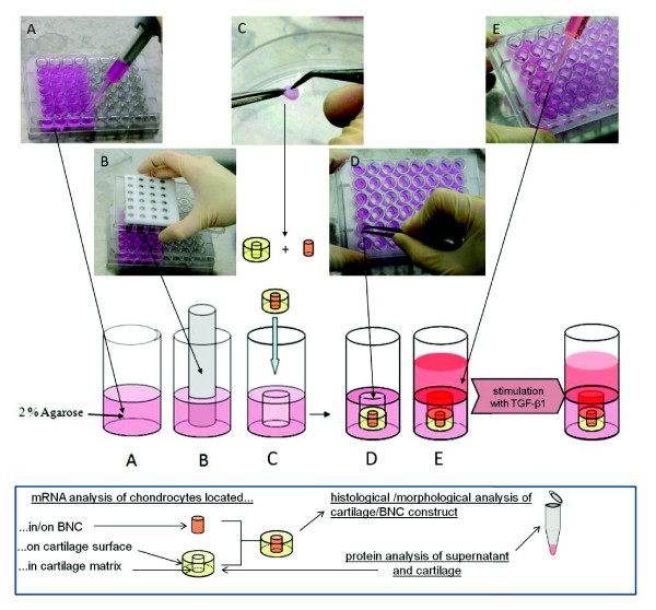
Current therapies for articular cartilage defects fail to achieve qualitatively sufficient tissue regeneration, possibly because of a mismatch between the speed of cartilage rebuilding and the resorption of degradable implant polymers. The present study focused on the self-healing capacity of resident cartilage cells in conjunction with cell-free and biocompatible (but non-resorbable) bacterial nanocellulose (BNC). This was tested in a novel in vitro bovine cartilage punch model.
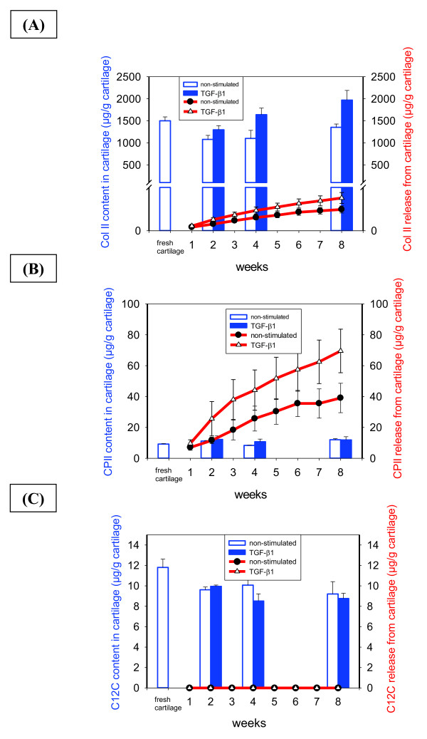
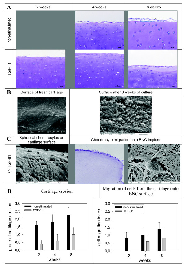
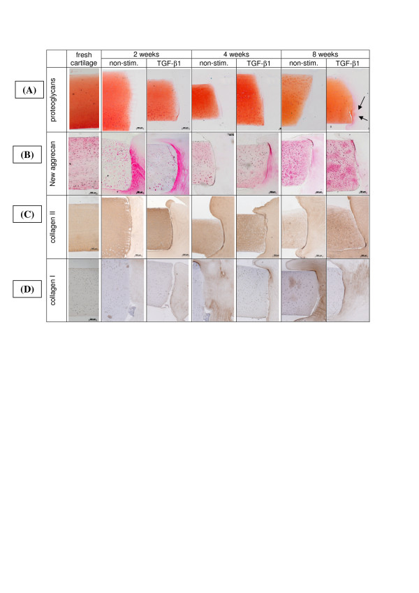
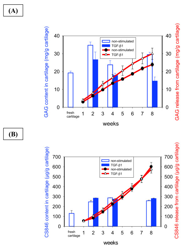
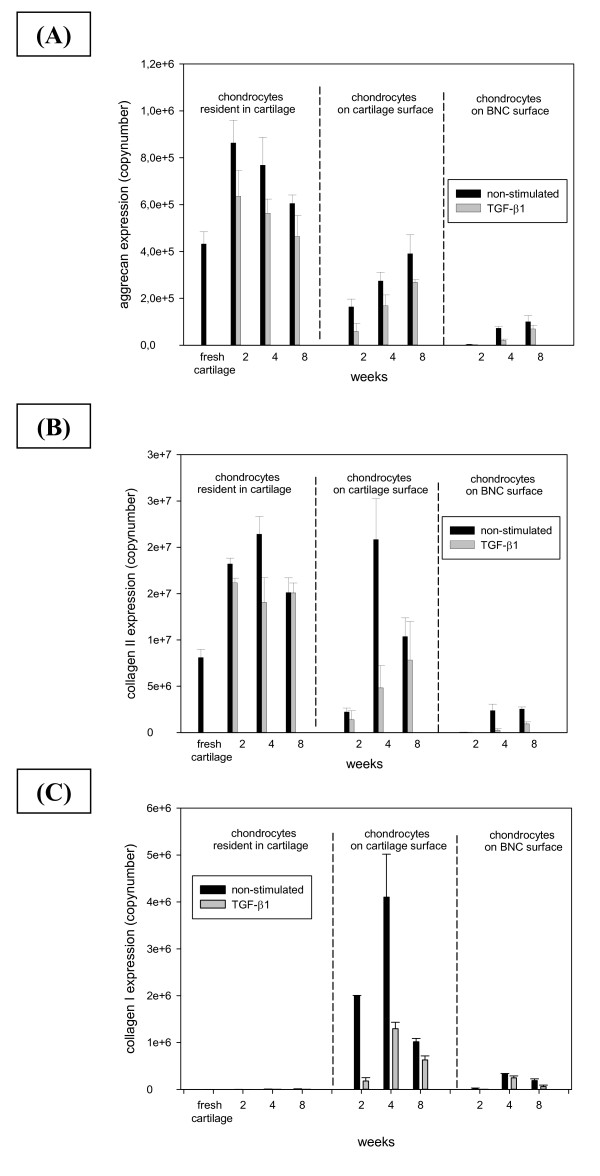
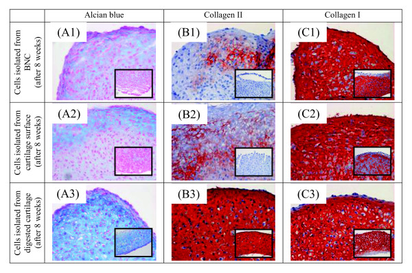
Standardized bovine cartilage discs with a central defect filled with BNC were cultured for up to eight weeks with/without stimulation with transforming growth factor-β1 (TGF-β1. Cartilage formation and integrity were analyzed by histology, immunohistochemistry and electron microscopy. Content, release and neosynthesis of the matrix molecules proteoglycan/aggrecan, collagen II and collagen I were also quantified. Finally, gene expression of these molecules was profiled in resident chondrocytes and chondrocytes migrated onto the cartilage surface or the implant material.
Non-stimulated and especially TGF-β1-stimulated cartilage discs displayed a preserved structural and functional integrity of the chondrocytes and surrounding matrix, remained vital in long-term culture (eight weeks) without signs of degeneration and showed substantial synthesis of cartilage-specific molecules at the protein and mRNA level. Whereas mobilization of chondrocytes from the matrix onto the surface of cartilage and implant was pivotal for successful seeding of cell-free BNC, chondrocytes did not immigrate into the central BNC area, possibly due to the relatively small diameter of its pores (2 to 5 μm). Chondrocytes on the BNC surface showed signs of successful redifferentiation over time, including increase of aggrecan/collagen type II mRNA, decrease of collagen type I mRNA and initial deposition of proteoglycan and collagen type II in long-term high-density pellet cultures. Although TGF-β1 stimulation showed protective effects on matrix integrity, effects on other parameters were limited.
The present bovine cartilage punch model represents a robust, reproducible and highly suitable tool for the long-term culture of cartilage, maintaining matrix integrity and homoeostasis. As an alternative to animal studies, this model may closely reflect early stages of cartilage regeneration, allowing the evaluation of promising biomaterials with/without chondrogenic factors.
目前用于治疗关节软骨缺损的疗法未能实现质量上足够的组织再生,这可能是因为软骨重建速度与可降解植入聚合物的吸收之间存在不匹配。本研究聚焦于驻留软骨细胞与无细胞且生物相容(但不可吸收)的细菌纳米纤维素(BNC)相结合的自我修复能力。这在一种新型的体外牛软骨打孔模型中进行了测试。
将中央缺损填充有BNC的标准化牛软骨圆盘在有/无转化生长因子-β1(TGF-β1)刺激的情况下培养长达8周。通过组织学、免疫组织化学和电子显微镜分析软骨形成和完整性。还对基质分子蛋白聚糖/聚集蛋白聚糖、胶原蛋白II和胶原蛋白I的含量、释放和新合成进行了定量。最后,在驻留软骨细胞以及迁移到软骨表面或植入材料上的软骨细胞中对这些分子的基因表达进行了分析。
未受刺激以及尤其是受TGF-β1刺激的软骨圆盘显示软骨细胞和周围基质的结构和功能完整性得以保留,在长期培养(8周)中保持活力且无退化迹象,并在蛋白质和mRNA水平上显示出大量软骨特异性分子的合成。虽然软骨细胞从基质向软骨和植入物表面的迁移对于无细胞BNC的成功接种至关重要,但软骨细胞并未迁移到中央BNC区域,这可能是由于其孔隙直径相对较小(2至5μm)。随着时间的推移,BNC表面的软骨细胞显示出成功再分化的迹象,包括聚集蛋白聚糖/II型胶原蛋白mRNA增加、I型胶原蛋白mRNA减少以及在长期高密度微球培养中蛋白聚糖和II型胶原蛋白的初始沉积。虽然TGF-β1刺激对基质完整性显示出保护作用,但其对其他参数的影响有限。
目前的牛软骨打孔模型是用于软骨长期培养、维持基质完整性和内环境稳定的一种强大、可重复且非常合适的工具。作为动物研究的替代方法,该模型可以密切反映软骨再生的早期阶段,从而能够评估有前景的生物材料以及是否添加软骨形成因子的情况。