Pedersen Torbjorn O, Blois Anna L, Xing Zhe, Xue Ying, Sun Yang, Finne-Wistrand Anna, Akslen Lars A, Lorens James B, Leknes Knut N, Fristad Inge, Mustafa Kamal
Stem Cell Res Ther. 2013 May 17;4(3):52. doi: 10.1186/scrt202.
A major determinant of the potential size of cell/scaffold constructs in tissue engineering is vascularization. The aims of this study were twofold: first to determine the in vitro angiogenic and osteogenic gene-expression profiles of endothelial cells (ECs) and mesenchymal stem cells (MSCs) cocultured in a dynamic 3D environment; and second, to assess differentiation and the potential for osteogenesis after in vivo implantation.
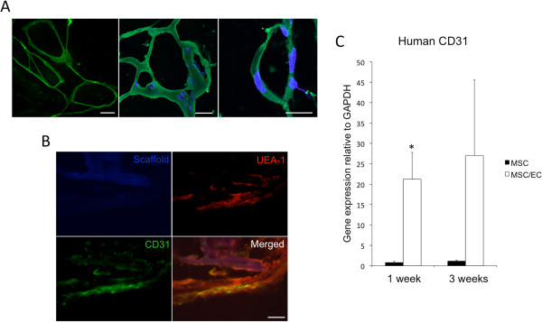
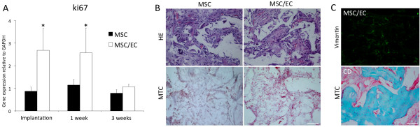
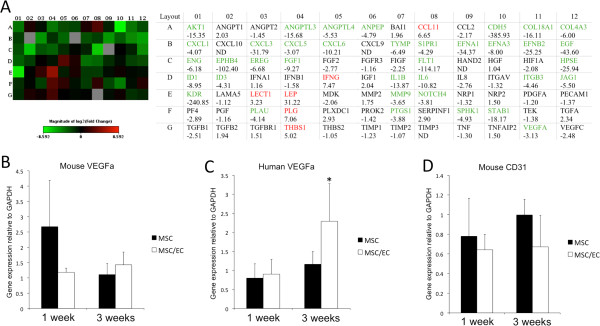
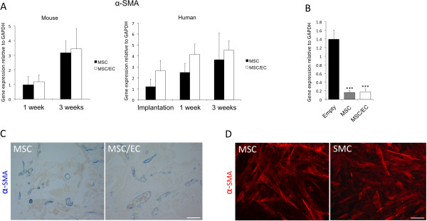
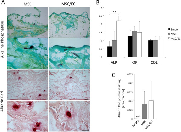
MSCs and ECs were grown in dynamic culture in poly(L-lactide-co-1,5-dioxepan-2-one) (poly(LLA-co-DXO)) copolymer scaffolds for 1 week, to generate three-dimensional endothelial microvascular networks. The constructs were then implanted in vivo, in a murine model for ectopic bone formation. Expression of selected genes for angiogenesis and osteogenesis was studied after a 1-week culture in vitro. Human cell proliferation was assessed as expression of ki67, whereas α-smooth muscle actin was used to determine the perivascular differentiation of MSCs. Osteogenesis was evaluated in vivo through detection of selected markers, by using real-time RT-PCR, alkaline phosphatase (ALP), Alizarin Red, hematoxylin/eosin (HE), and Masson trichrome staining.
The results show that endothelial microvascular networks could be generated in a poly(LLA-co-DXO) scaffold in vitro and sustained after in vivo implantation. The addition of ECs to MSCs influenced both angiogenic and osteogenic gene-expression profiles. Furthermore, human ki67 was upregulated before and after implantation. MSCs could support functional blood vessels as perivascular cells independent of implanted ECs. In addition, the expression of ALP was upregulated in the presence of endothelial microvascular networks.
This study demonstrates that copolymer poly(LLA-co-DXO) scaffolds can be prevascularized with ECs and MSCs. Although a local osteoinductive environment is required to achieve ectopic bone formation, seeding of MSCs with or without ECs increases the osteogenic potential of tissue-engineered constructs.
组织工程中细胞/支架构建体潜在大小的一个主要决定因素是血管化。本研究有两个目的:一是确定在动态三维环境中共培养的内皮细胞(ECs)和间充质干细胞(MSCs)的体外血管生成和成骨基因表达谱;二是评估体内植入后的分化和成骨潜力。
将MSCs和ECs在聚(L-丙交酯-co-1,5-二氧杂环庚烷-2-酮)(聚(LLA-co-DXO))共聚物支架中进行动态培养1周,以生成三维内皮微血管网络。然后将构建体植入体内,用于异位骨形成的小鼠模型。在体外培养1周后研究血管生成和成骨相关选定基因的表达。通过检测ki67的表达评估人细胞增殖,而α-平滑肌肌动蛋白用于确定MSCs的血管周分化。通过实时逆转录聚合酶链反应、碱性磷酸酶(ALP)、茜素红、苏木精/伊红(HE)和Masson三色染色检测选定标志物,在体内评估成骨情况。
结果表明,体外可在聚(LLA-co-DXO)支架中生成内皮微血管网络,并在体内植入后得以维持。将ECs添加到MSCs中会影响血管生成和成骨基因表达谱。此外,人ki67在植入前后均上调。MSCs可作为血管周细胞支持功能性血管,独立于植入的ECs。此外,在内皮微血管网络存在的情况下,ALP的表达上调。
本研究表明,共聚物聚(LLA-co-DXO)支架可通过ECs和MSCs进行预血管化。尽管需要局部骨诱导环境来实现异位骨形成,但接种有或没有ECs的MSCs可增加组织工程构建体的成骨潜力。