• 文献检索
  • 文档翻译
  • 深度研究
  • 学术资讯
  • Suppr Zotero 插件Zotero 插件
  • 邀请有礼
  • 套餐&价格
  • 历史记录
应用&插件
Suppr Zotero 插件Zotero 插件浏览器插件Mac 客户端Windows 客户端微信小程序
定价
高级版会员购买积分包购买API积分包
服务
文献检索文档翻译深度研究API 文档MCP 服务
关于我们
关于 Suppr公司介绍联系我们用户协议隐私条款
关注我们

Suppr 超能文献

核心技术专利:CN118964589B侵权必究
粤ICP备2023148730 号-1Suppr @ 2026

文献检索

告别复杂PubMed语法,用中文像聊天一样搜索,搜遍4000万医学文献。AI智能推荐,让科研检索更轻松。

立即免费搜索

文件翻译

保留排版,准确专业,支持PDF/Word/PPT等文件格式,支持 12+语言互译。

免费翻译文档

深度研究

AI帮你快速写综述,25分钟生成高质量综述,智能提取关键信息,辅助科研写作。

立即免费体验

仿生细胞支架共培养 MSC 源性成骨和成血管细胞增强骨组织再生。

Enhanced bone tissue regeneration of a biomimetic cellular scaffold with co-cultured MSCs-derived osteogenic and angiogenic cells.

机构信息

Research Center for Nano-Biomaterials, Analytical & Testing Center, Sichuan University, Chengdu, China.

Technology Transfer Center, Kunming Medical University, Kunming, China.

出版信息

Cell Prolif. 2019 Sep;52(5):e12658. doi: 10.1111/cpr.12658. Epub 2019 Jul 11.

DOI:10.1111/cpr.12658
PMID:31297910
原文链接:https://pmc.ncbi.nlm.nih.gov/articles/PMC6797511/
Abstract

OBJECTIVES

The bone tissue engineering primarily focuses on three-dimensional co-culture systems, which physical and biological properties resemble the cell matrix of actual tissues. The complex dialogue between bone-forming and endothelial cells (ECs) in a tissue-engineered construct will directly regulate angiogenesis and bone regeneration. The purpose of this study was to investigate whether co-culture between osteogenic and angiogenic cells derived by bone mesenchymal stem cells (MSCs) could affect cell activities and new bone formation.

MATERIALS AND METHODS

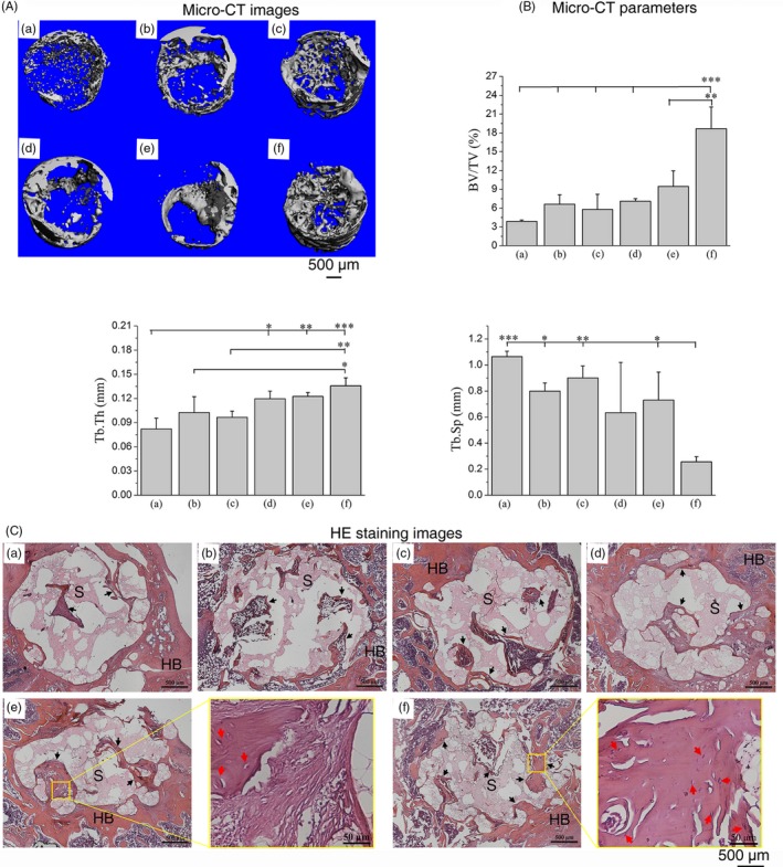
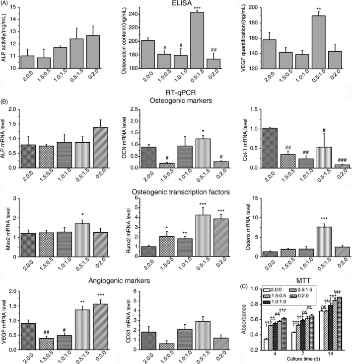
Mesenchymal stem cells were dually induced to differentiate into osteogenic cells (OMSCs) and ECs; both cell types were co-cultured at different ratios to investigate their effects and underlying mechanisms through ELISA, RT-qPCR and MTT assays. The selected cell mixture was transplanted onto a nano-hydroxyapatite/polyurethane (n-HA/PU) scaffold to form a cell-scaffold construct that was implanted in the rat femoral condyles. Histology and micro-CT were examined for further verification.

RESULTS

ELISA and gene expression studies revealed that co-cultured OMSCs/ECs (0.5/1.5) significantly elevated the transcription levels of osteogenic genes such as ALP, Col-I and OCN, as well as transcription factors Msx2, Runx2 and Osterix; it also upregulated angiogenic factors of vascular endothelial growth factor (VEGF) and CD31 when compared with cells cultured alone or in other ratios. The optimized OMSCs/ECs group had more abundant calcium phosphate crystal deposition, further facilitated their bone formation in vivo.

CONCLUSIONS

The OMSCs/ECs-scaffold constructs at an optimal cell ratio (0.5/1.5) achieved enhanced osteogenic and angiogenic factor expression and biomineralization, which resulted in more effective bone formation.

摘要

目的

骨组织工程主要侧重于三维共培养系统,其物理和生物特性类似于实际组织的细胞基质。在组织工程构建体中,成骨细胞和内皮细胞(ECs)之间的复杂对话将直接调节血管生成和骨再生。本研究旨在探讨成骨细胞和血管生成细胞是否可以通过骨髓间充质干细胞(MSCs)共培养来影响细胞活性和新骨形成。

材料和方法

将间充质干细胞双重诱导分化为成骨细胞(OMSCs)和 ECs;将两种细胞类型以不同比例共培养,通过 ELISA、RT-qPCR 和 MTT 检测来研究它们的作用和潜在机制。选择的细胞混合物被移植到纳米羟基磷灰石/聚氨酯(n-HA/PU)支架上,形成细胞-支架构建体,然后植入大鼠股骨髁。通过组织学和 micro-CT 进行进一步验证。

结果

ELISA 和基因表达研究表明,共培养的 OMSCs/ECs(0.5/1.5)显著提高了碱性磷酸酶(ALP)、Col-I 和 OCN 等成骨基因以及 Msx2、Runx2 和 Osterix 等转录因子的转录水平;与单独培养或其他比例培养的细胞相比,它还上调了血管内皮生长因子(VEGF)和 CD31 等血管生成因子。优化的 OMSCs/ECs 组具有更丰富的钙磷晶体沉积,进一步促进了其体内成骨作用。

结论

最佳细胞比例(0.5/1.5)的 OMSCs/ECs-支架构建体实现了增强的成骨和血管生成因子表达和生物矿化,从而更有效地促进了骨形成。

https://cdn.ncbi.nlm.nih.gov/pmc/blobs/e568/6797511/8bbd7b8ee4cf/CPR-52-e12658-g005.jpg
https://cdn.ncbi.nlm.nih.gov/pmc/blobs/e568/6797511/151797a249d7/CPR-52-e12658-g001.jpg
https://cdn.ncbi.nlm.nih.gov/pmc/blobs/e568/6797511/c41926548720/CPR-52-e12658-g002.jpg
https://cdn.ncbi.nlm.nih.gov/pmc/blobs/e568/6797511/301df78b4dfb/CPR-52-e12658-g003.jpg
https://cdn.ncbi.nlm.nih.gov/pmc/blobs/e568/6797511/415a2f82af91/CPR-52-e12658-g004.jpg
https://cdn.ncbi.nlm.nih.gov/pmc/blobs/e568/6797511/8bbd7b8ee4cf/CPR-52-e12658-g005.jpg

相似文献

1
Enhanced bone tissue regeneration of a biomimetic cellular scaffold with co-cultured MSCs-derived osteogenic and angiogenic cells.仿生细胞支架共培养 MSC 源性成骨和成血管细胞增强骨组织再生。
Cell Prolif. 2019 Sep;52(5):e12658. doi: 10.1111/cpr.12658. Epub 2019 Jul 11.
2
Osteogenic differentiation and bone regeneration of iPSC-MSCs supported by a biomimetic nanofibrous scaffold.仿生纳米纤维支架支持的诱导多能干细胞来源间充质干细胞的成骨分化与骨再生
Acta Biomater. 2016 Jan;29:365-379. doi: 10.1016/j.actbio.2015.10.007. Epub 2015 Oct 9.
3
Sema3A and HIF1α co-overexpressed iPSC-MSCs/HA scaffold facilitates the repair of calvarial defect in a mouse model.Sema3A 和 HIF1α 共过表达 iPSC-MSCs/HA 支架促进了小鼠颅骨缺损模型的修复。
J Cell Physiol. 2020 Oct;235(10):6754-6766. doi: 10.1002/jcp.29569. Epub 2020 Feb 3.
4
A novel method to improve the osteogenesis capacity of hUCMSCs with dual-directional pre-induction under screened co-culture conditions.一种在筛选共培养条件下通过双向预诱导提高人 UCMSCs 成骨能力的新方法。
Cell Prolif. 2020 Feb;53(2):e12740. doi: 10.1111/cpr.12740. Epub 2019 Dec 9.
5
Endothelial microvascular networks affect gene-expression profiles and osteogenic potential of tissue-engineered constructs.内皮微血管网络影响组织工程构建体的基因表达谱和成骨潜力。
Stem Cell Res Ther. 2013 May 17;4(3):52. doi: 10.1186/scrt202.
6
Angiogenic and osteogenic regeneration in rats via calcium phosphate scaffold and endothelial cell co-culture with human bone marrow mesenchymal stem cells (MSCs), human umbilical cord MSCs, human induced pluripotent stem cell-derived MSCs and human embryonic stem cell-derived MSCs.通过磷酸钙支架和人骨髓间充质干细胞(MSCs)、人脐带 MSCs、人诱导多能干细胞源性 MSCs 和人胚胎干细胞源性 MSCs 与内皮细胞共培养,在大鼠中实现血管生成和成骨再生。
J Tissue Eng Regen Med. 2018 Jan;12(1):191-203. doi: 10.1002/term.2395. Epub 2017 Jun 13.
7
[A novel tissue-engineered bone constructed by using human adipose-derived stem cells and biomimetic calcium phosphate scaffold coprecipitated with bone morphogenetic protein-2].一种通过使用人脂肪来源干细胞和与骨形态发生蛋白-2共沉淀的仿生磷酸钙支架构建的新型组织工程骨
Beijing Da Xue Xue Bao Yi Xue Ban. 2017 Feb 18;49(1):6-15.
8
Electrospun biomimetic scaffold of hydroxyapatite/chitosan supports enhanced osteogenic differentiation of mMSCs.电纺仿生羟基磷灰石/壳聚糖支架促进骨髓间充质干细胞的成骨分化。
Nanotechnology. 2012 Dec 7;23(48):485102. doi: 10.1088/0957-4484/23/48/485102. Epub 2012 Nov 6.
9
Naringin-inlaid silk fibroin/hydroxyapatite scaffold enhances human umbilical cord-derived mesenchymal stem cell-based bone regeneration.柚皮苷镶嵌丝素蛋白/羟基磷灰石支架增强人脐带间充质干细胞的成骨再生。
Cell Prolif. 2021 Jul;54(7):e13043. doi: 10.1111/cpr.13043. Epub 2021 May 19.
10
Small molecules modified biomimetic gelatin/hydroxyapatite nanofibers constructing an ideal osteogenic microenvironment with significantly enhanced cranial bone formation.小分子修饰仿生明胶/羟基磷灰石纳米纤维构建理想的成骨微环境,显著增强颅骨骨形成。
Int J Nanomedicine. 2018 Nov 6;13:7167-7181. doi: 10.2147/IJN.S174553. eCollection 2018.

引用本文的文献

1
Enhancing bone regeneration using kaempferol as an osteoprotective compound: signaling mechanisms, delivery strategies, and potential applications.使用山奈酚作为骨保护化合物增强骨再生:信号传导机制、递送策略及潜在应用
J Biol Eng. 2025 Aug 6;19(1):74. doi: 10.1186/s13036-025-00545-5.
2
Strategies for promoting neurovascularization in bone regeneration.促进骨再生中神经血管化的策略。
Mil Med Res. 2025 Mar 3;12(1):9. doi: 10.1186/s40779-025-00596-1.
3
Analysis of the Effect of Human Type I Collagen-Derived Peptide on Bone Regenerative Capacity and Comparison with Various Collagen Materials In Vivo.

本文引用的文献

1
An injectable double-network hydrogel for the co-culture of vascular endothelial cells and bone marrow mesenchymal stem cells for simultaneously enhancing vascularization and osteogenesis.一种用于血管内皮细胞和骨髓间充质干细胞共培养的可注射双网络水凝胶,可同时增强血管生成和成骨作用。
J Mater Chem B. 2018 Dec 21;6(47):7811-7821. doi: 10.1039/c8tb02244e. Epub 2018 Oct 22.
2
TiO₂ Nanotubes Enhance Vascularization and Osteogenic Differentiation Through Stimulating Interactions Between Bone Marrow Stromal Cells and Endothelial Cells.TiO₂ 纳米管通过刺激骨髓基质细胞和内皮细胞之间的相互作用来增强血管生成和成骨分化。
J Biomed Nanotechnol. 2018 Apr 1;14(4):765-777. doi: 10.1166/jbn.2018.2508.
3
人I型胶原衍生肽对骨再生能力的影响分析及其与多种胶原材料的体内比较。
Medicina (Kaunas). 2025 Jan 2;61(1):57. doi: 10.3390/medicina61010057.
4
Revolutionizing bone defect healing: the power of mesenchymal stem cells as seeds.革新骨缺损愈合:间充质干细胞作为种子的力量。
Front Bioeng Biotechnol. 2024 Oct 21;12:1421674. doi: 10.3389/fbioe.2024.1421674. eCollection 2024.
5
Combining "waste utilization" and "tissue to tissue" strategies to accelerate vascularization for bone repair.结合“废物利用”和“组织对组织”策略以加速骨修复的血管化
J Orthop Translat. 2024 Jun 24;47:132-143. doi: 10.1016/j.jot.2024.04.002. eCollection 2024 Jul.
6
Towards Stem Cell Therapy for Critical-Sized Segmental Bone Defects: Current Trends and Challenges on the Path to Clinical Translation.走向用于大段临界尺寸骨缺损的干细胞治疗:临床转化之路的当前趋势与挑战
J Funct Biomater. 2024 May 27;15(6):145. doi: 10.3390/jfb15060145.
7
Biological Effects, Applications and Design Strategies of Medical Polyurethanes Modified by Nanomaterials.纳米材料改性医用聚氨酯的生物效应、应用及设计策略。
Int J Nanomedicine. 2022 Dec 29;17:6791-6819. doi: 10.2147/IJN.S393207. eCollection 2022.
8
An injectable pH neutral bioactive glass-based bone cement with suitable bone regeneration ability.一种具有适宜骨再生能力的可注射pH中性生物活性玻璃基骨水泥。
J Orthop Translat. 2022 Sep 2;36:120-131. doi: 10.1016/j.jot.2022.05.011. eCollection 2022 Sep.
9
Sequential gastrodin release PU/n-HA composite scaffolds reprogram macrophages for improved osteogenesis and angiogenesis.序贯释放天麻素的PU/n-HA复合支架可重编程巨噬细胞以促进骨生成和血管生成。
Bioact Mater. 2022 Apr 1;19:24-37. doi: 10.1016/j.bioactmat.2022.03.037. eCollection 2023 Jan.
10
Bone Regeneration and Oxidative Stress: An Updated Overview.骨再生与氧化应激:最新综述
Antioxidants (Basel). 2022 Feb 6;11(2):318. doi: 10.3390/antiox11020318.
In vitro interaction of human Wharton's jelly mesenchymal stem cells with biomimetic 3D scaffold.
人脐带华通氏胶间充质干细胞与仿生 3D 支架的体外相互作用。
J Biomed Mater Res A. 2019 Jun;107(6):1166-1175. doi: 10.1002/jbm.a.36608. Epub 2019 Mar 18.
4
The effects of biomimetically conjugated VEGF on osteogenesis and angiogenesis of MSCs (human and rat) and HUVECs co-culture models.仿生连接的 VEGF 对人源和鼠源 MSCs 与 HUVECs 共培养模型中成骨和血管生成的影响。
Colloids Surf B Biointerfaces. 2018 Jul 1;167:550-559. doi: 10.1016/j.colsurfb.2018.04.060. Epub 2018 Apr 30.
5
Tension-loaded bone marrow stromal cells potentiate the paracrine osteogenic signaling of co-cultured vascular endothelial cells.张力加载的骨髓基质细胞增强共培养血管内皮细胞的旁分泌成骨信号。
Biol Open. 2018 Jun 13;7(6):bio032482. doi: 10.1242/bio.032482.
6
Vitamin D Promotes MSC Osteogenic Differentiation Stimulating Cell Adhesion and V3 Expression.维生素D通过刺激细胞黏附和V3表达促进间充质干细胞成骨分化。
Stem Cells Int. 2018 Feb 28;2018:6958713. doi: 10.1155/2018/6958713. eCollection 2018.
7
Glucosamine-modified polyethylene glycol hydrogel-mediated chondrogenic differentiation of human mesenchymal stem cells.葡萄糖胺修饰的聚乙二醇水凝胶介导人间充质干细胞的软骨形成分化
Mater Sci Eng C Mater Biol Appl. 2017 Oct 1;79:661-670. doi: 10.1016/j.msec.2017.05.043. Epub 2017 May 11.
8
Co-Seeding Human Endothelial Cells with Human-Induced Pluripotent Stem Cell-Derived Mesenchymal Stem Cells on Calcium Phosphate Scaffold Enhances Osteogenesis and Vascularization in Rats.在磷酸钙支架上共同接种人诱导多能干细胞衍生的间充质干细胞与人内皮细胞可增强大鼠的成骨作用和血管生成。
Tissue Eng Part A. 2017 Jun;23(11-12):546-555. doi: 10.1089/ten.tea.2016.0485. Epub 2017 Mar 10.
9
Repair of rat critical size calvarial defect using osteoblast-like and umbilical vein endothelial cells seeded in gelatin/hydroxyapatite scaffolds.使用接种于明胶/羟基磷灰石支架的成骨样细胞和脐静脉内皮细胞修复大鼠临界尺寸颅骨缺损。
J Biomed Mater Res A. 2016 Jul;104(7):1770-8. doi: 10.1002/jbm.a.35710. Epub 2016 Mar 21.
10
Oxygen Tension-Controlled Matrices with Osteogenic and Vasculogenic Cells for Vascularized Bone Regeneration In Vivo.用于体内血管化骨再生的含成骨细胞和血管生成细胞的氧张力控制基质
Tissue Eng Part A. 2016 Apr;22(7-8):610-20. doi: 10.1089/ten.TEA.2015.0310. Epub 2016 Mar 22.