Department of Radiology, School of Medicine, Ewha Womans University, Seoul 158-710, Korea.
Korean J Radiol. 2013 May-Jun;14(3):532-9. doi: 10.3348/kjr.2013.14.3.532. Epub 2013 May 2.
To evaluate the reliability of virtual non-contrast (VNC) images reconstructed from contrast-enhanced, dual-energy scans compared with true non-contrast (TNC) images in the assessment of high CT attenuation or calcification of mediastinal lymph nodes.
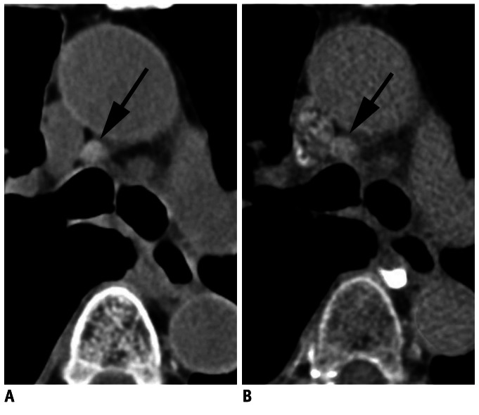
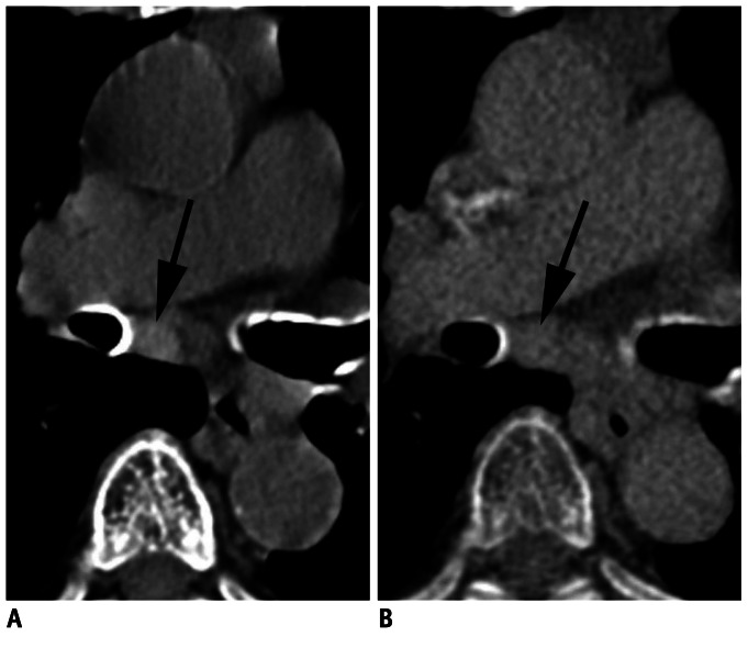
A total of 112 mediastinal nodes from 45 patients who underwent non-contrast and dual-energy contrast-enhanced scans were analyzed. Node attenuation in TNC and VNC images was compared both objectively, using computed tomography (CT) attenuation, and subjectively, via visual scoring (0, attenuation ≤ the aorta; 1, > the aorta; 2, calcification). The relationship among attenuation difference between TNC and VNC images, CT attenuation in TNC images, and net contrast enhancement (NCE) was analyzed.
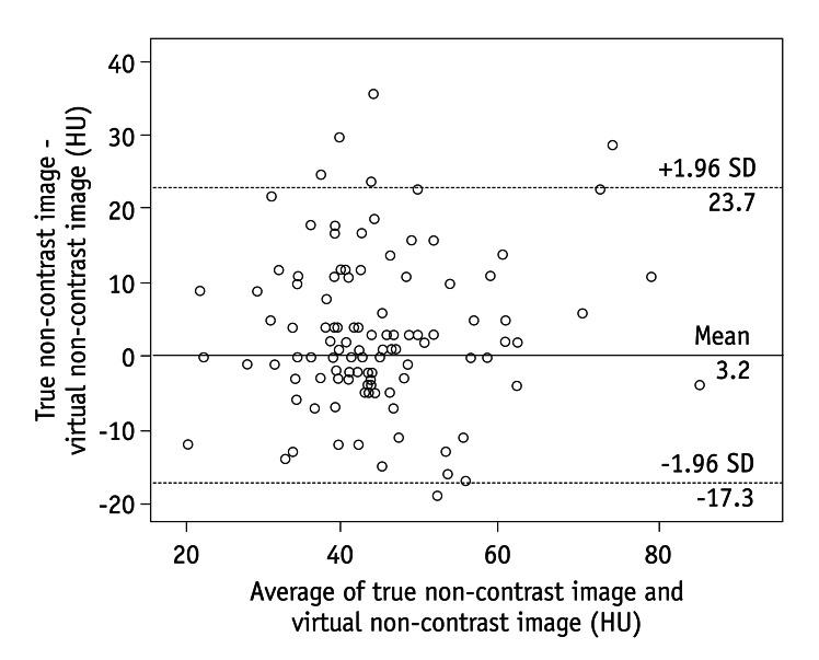
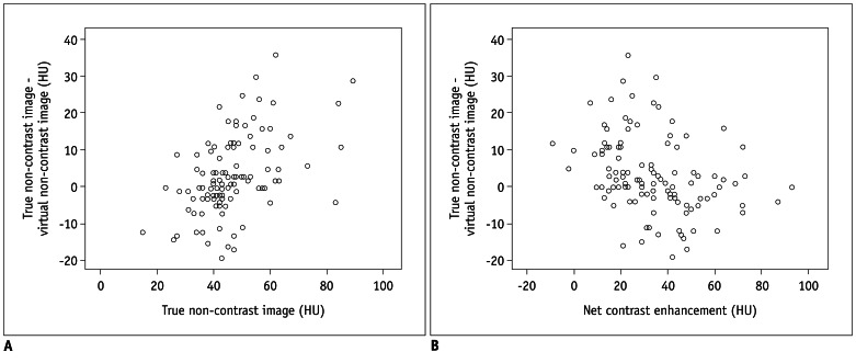
CT attenuation in TNC and VNC images showed moderate agreement (intraclass correlation coefficient, 0.612). The mean absolute difference was 7.8 ± 7.6 Hounsfield unit (HU) (range, 0-36 HU), and the absolute difference was equal to or less than 10 HU in 65.2% of cases (73/112). Visual scores in TNC and VNC images showed fair agreement (κ value, 0.335). Five of 16 nodes (31.3%) which showed score 1 (n = 15) or 2 (n = 1) in TNC images demonstrated score 1 in VNC images. The TNC-VNC attenuation difference showed a moderate positive correlation with CT attenuation in TNC images (partial correlation coefficient [PCC] adjusted by NCE: 0.455) and a weak negative correlation with NCE (PCC adjusted by CT attenuation in TNC: -0.245).
VNC images may be useful in the evaluation of mediastinal lymph nodes by providing additional information of high CT attenuation of nodes, although it is underestimated compared with TNC images.
评估对比增强双能扫描重建的虚拟非对比(VNC)图像与真实非对比(TNC)图像在评估纵隔淋巴结高 CT 衰减或钙化中的可靠性。
共分析了 45 例接受非对比和双能对比增强扫描的患者的 112 个纵隔淋巴结。分别从客观和主观两个方面比较了 TNC 和 VNC 图像中淋巴结的衰减情况,客观方面采用 CT 衰减值,主观方面采用视觉评分(0:衰减值≤主动脉;1:>主动脉;2:钙化)。分析了 TNC 和 VNC 图像之间的衰减差异与 TNC 图像 CT 衰减值和净对比增强(NCE)之间的关系。
TNC 和 VNC 图像的 CT 衰减值具有中度一致性(组内相关系数,0.612)。平均绝对差值为 7.8±7.6HU(范围:0-36HU),差值在 10HU 以内的比例为 65.2%(73/112)。TNC 和 VNC 图像的视觉评分显示出适度的一致性(κ 值,0.335)。在 TNC 图像中评分 1(n=15)或 2(n=1)的 16 个淋巴结中,有 5 个(31.3%)在 VNC 图像中也表现为评分 1。TNC-VNC 衰减差值与 TNC 图像 CT 衰减值呈中度正相关(经 NCE 校正的部分相关系数[PCC]:0.455),与 NCE 呈弱负相关(经 TNC 图像 CT 衰减值校正的 PCC:-0.245)。
尽管与 TNC 图像相比,VNC 图像低估了纵隔淋巴结的 CT 衰减值,但它可以提供淋巴结高 CT 衰减的额外信息,因此在评估纵隔淋巴结方面可能是有用的。