Department of Physiology, Chonnam National University Medical School, Gwangju, Korea.
PLoS One. 2013 May 17;8(5):e63186. doi: 10.1371/journal.pone.0063186. Print 2013.
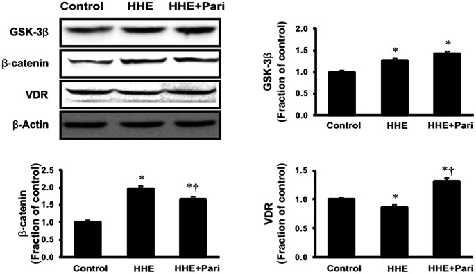
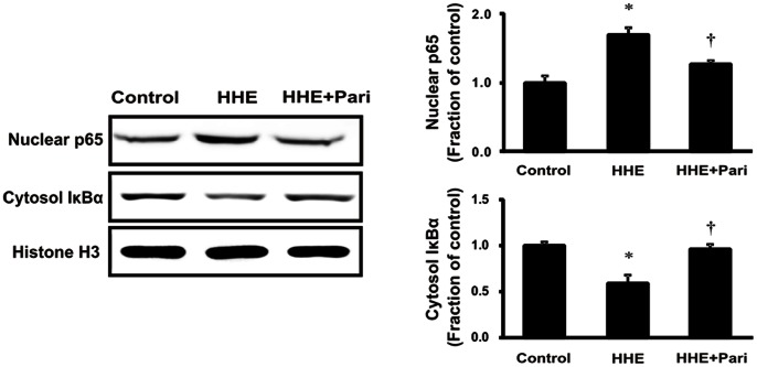
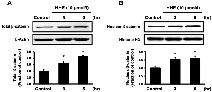
4-Hydroxy-2-hexenal (HHE), the aldehyde product of lipid peroxidation, may be responsible for the pathogenesis of progressive renal disease. Recently, paricalcitol (19-nor-1,25-dihydroxyvitamin D2) was shown to be renoprotective through its anti-inflammatory and antifibrotic effects in various experimental nephropathy models. In this study, we investigated the effects of paricalcitol on inflammation and epithelial-mesenchymal transition (EMT) after HHE-induced renal tubular epithelial cell injury. To investigate the molecular mechanisms underlying HHE-induced renal tubular cell injury, the human proximal tubular epithelial (HK-2) cells cultured with 10 µM HHE in the presence or absence of paricalcitol. In HK-2 cells, paricalcitol attenuated the HHE-induced expression of extracellular signal-regulated kinase, c-Jun N-terminal kinase, and p38 mitogen-activated protein kinase, and prevented nuclear factor-κB (NF-κB) activation. The expression of the inflammatory proteins inducible nitric oxide synthase and cyclooxygenase-2 was attenuated by paricalcitol pretreatment. In addition, HHE increased the expression of the transforming growth factor (TGF)-β/Smad signaling proteins and fibrotic proteins, such as α-smooth muscle actin and connective tissue growth factor; this inducible expression was suppressed by pretreatment with paricalcitol. Treatment with HHE resulted in the activation of the β-catenin signaling pathway, and paricalcitol pretreatment reduced the expression of β-catenin in HHE-treated HK-2 cells. Coimmunoprecipitation shows that paricalcitol induced vitamin D receptor (VDR)/β-catenin complex formation in HK-2 cells. Also immunofluorescence staining revealed that co-localization of VDR and β-catenin in the nuclei. ICG-001, an inhibitor of β-catenin, decreased the expression of TGF-β1 and attenuated HHE-induced tubular EMT. These results show that paricalcitol attenuated HHE-induced renal tubular cell injury by suppressing inflammation and EMT process through inhibition of the NF-κB, TGF-β/Smad, and β-catenin signaling pathways.
4-羟基-2-己烯醛(HHE),脂质过氧化的醛产物,可能是进行性肾病发病机制的罪魁祸首。最近,研究表明,帕立骨化醇(19-去甲-1,25-二羟维生素 D2)通过其在各种实验性肾病模型中的抗炎和抗纤维化作用具有肾脏保护作用。在这项研究中,我们研究了帕立骨化醇对 HHE 诱导的肾小管上皮细胞损伤后炎症和上皮-间充质转化(EMT)的影响。为了研究 HHE 诱导的肾小管细胞损伤的分子机制,我们在存在或不存在帕立骨化醇的情况下,用 10µM HHE 培养人近端肾小管上皮(HK-2)细胞。在 HK-2 细胞中,帕立骨化醇减弱了 HHE 诱导的细胞外信号调节激酶、c-Jun N-末端激酶和 p38 丝裂原活化蛋白激酶的表达,并阻止了核因子-κB(NF-κB)的激活。诱导型一氧化氮合酶和环氧化酶-2 的表达被帕立骨化醇预处理所减弱。此外,HHE 增加了转化生长因子(TGF)-β/Smad 信号蛋白和纤维蛋白的表达,如α-平滑肌肌动蛋白和结缔组织生长因子;这种诱导表达被帕立骨化醇预处理所抑制。用 HHE 处理导致β-连环蛋白信号通路的激活,而帕立骨化醇预处理减少了 HHE 处理的 HK-2 细胞中β-连环蛋白的表达。共免疫沉淀显示帕立骨化醇诱导 HK-2 细胞中维生素 D 受体(VDR)/β-连环蛋白复合物的形成。免疫荧光染色也显示 VDR 和β-连环蛋白在核内的共定位。β-连环蛋白抑制剂 ICG-001 降低了 TGF-β1 的表达,并减弱了 HHE 诱导的管状 EMT。这些结果表明,帕立骨化醇通过抑制 NF-κB、TGF-β/Smad 和β-连环蛋白信号通路,抑制炎症和 EMT 过程,减轻 HHE 诱导的肾小管细胞损伤。