Department of Pathology, University of Texas Medical Branch, Galveston, Texas, United States of America.
PLoS One. 2013 May 17;8(5):e63331. doi: 10.1371/journal.pone.0063331. Print 2013.
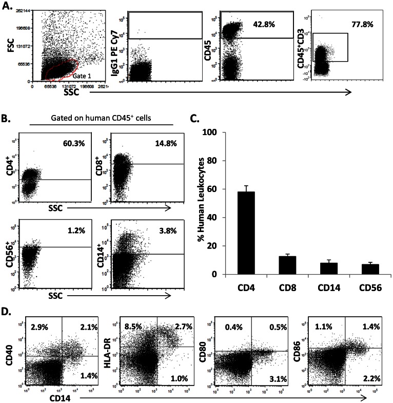
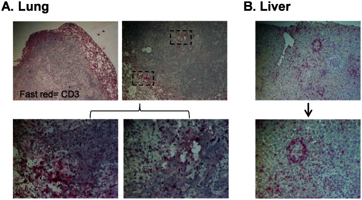
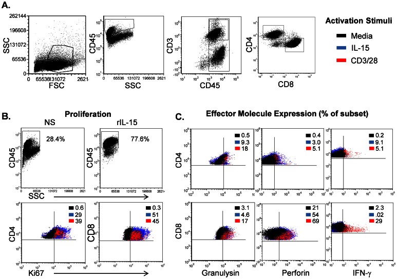
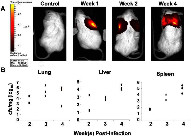
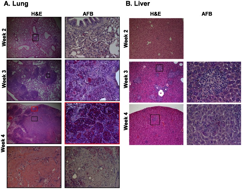
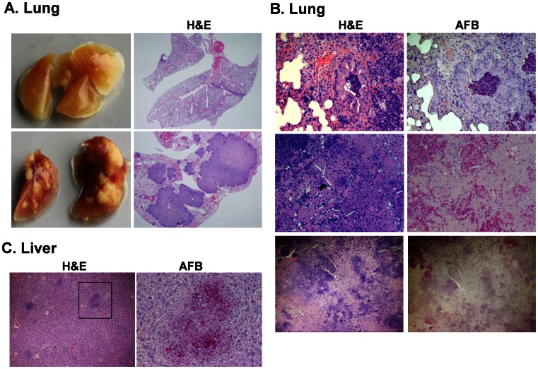
Mycobacterium tuberculosis (M.tb) is the second leading infectious cause of death worldwide and the primary cause of death in people living with HIV/AIDS. There are several excellent animal models employed to study tuberculosis (TB), but many have limitations for reproducing human pathology and none are amenable to the direct study of HIV/M.tb co-infection. The humanized mouse has been increasingly employed to explore HIV infection and other pathogens where animal models are limiting. Our goal was to develop a small animal model of M.tb infection using the bone marrow, liver, thymus (BLT) humanized mouse. NOD-SCID/γc(null) mice were engrafted with human fetal liver and thymus tissue, and supplemented with CD34(+) fetal liver cells. Excellent reconstitution, as measured by expression of the human CD45 pan leukocyte marker by peripheral blood populations, was observed at 12 weeks after engraftment. Human T cells (CD3, CD4, CD8), as well as natural killer cells and monocyte/macrophages were all observed within the human leukocyte (CD45(+)) population. Importantly, human T cells were functionally competent as determined by proliferative capacity and effector molecule (e.g. IFN-γ, granulysin, perforin) expression in response to positive stimuli. Animals infected intranasally with M.tb had progressive bacterial infection in the lung and dissemination to spleen and liver from 2-8 weeks post infection. Sites of infection in the lung were characterized by the formation of organized granulomatous lesions, caseous necrosis, bronchial obstruction, and crystallization of cholesterol deposits. Human T cells were distributed throughout the lung, liver, and spleen at sites of inflammation and bacterial growth and were organized to the periphery of granulomas. These preliminary results demonstrate the potential to use the humanized mouse as a model of experimental TB.
结核分枝杆菌(M.tb)是全球第二大致死性传染病病因,也是艾滋病毒/艾滋病患者的主要死因。有几种优秀的动物模型被用于研究结核病(TB),但许多模型在复制人类病理学方面存在局限性,也没有一种模型适合直接研究人类艾滋病毒/结核分枝杆菌合并感染。人源化小鼠越来越多地被用于探索在动物模型中存在局限性的艾滋病毒感染和其他病原体。我们的目标是使用骨髓、肝脏、胸腺(BLT)人源化小鼠建立结核分枝杆菌感染的小动物模型。NOD-SCID/γc(null) 小鼠被移植了人胎儿肝脏和胸腺组织,并补充了 CD34(+) 胎儿肝脏细胞。在移植后 12 周,通过外周血人群中人类 CD45 泛白细胞标志物的表达观察到了极好的重建。人 T 细胞(CD3、CD4、CD8)以及自然杀伤细胞和单核细胞/巨噬细胞都存在于人类白细胞(CD45(+))群体中。重要的是,人 T 细胞具有功能能力,这是通过增殖能力和对阳性刺激的效应分子(例如 IFN-γ、颗粒酶、穿孔素)表达来确定的。经鼻感染结核分枝杆菌的动物在感染后 2-8 周肺部出现进行性细菌感染并播散至脾和肝。肺部感染部位的特征是形成有组织的肉芽肿病变、干酪样坏死、支气管阻塞和胆固醇沉积结晶。人类 T 细胞分布在肺部、肝脏和脾脏的炎症和细菌生长部位,并组织到肉芽肿的外围。这些初步结果表明,人源化小鼠有可能成为实验性结核病的模型。