1. Department of Developmental Biology, Faculty of Biological Sciences, Kharazmi University, Tehran, Iran.
Cell J. 2013 Spring;15(1):29-36. Epub 2013 May 5.
Embryonic cerebrospinal fluid (e-CSF) has an important role in development of embryonic and adult brain. Proteomic analysis suggests that this fluid has many morphogenes and cytokines that alter in time and space throughout embryonic life. The aim of this study was to evaluate the developmental effect of embryonic CSF on proliferation and differentiation of neuroprogenitor cells in different gestational age.
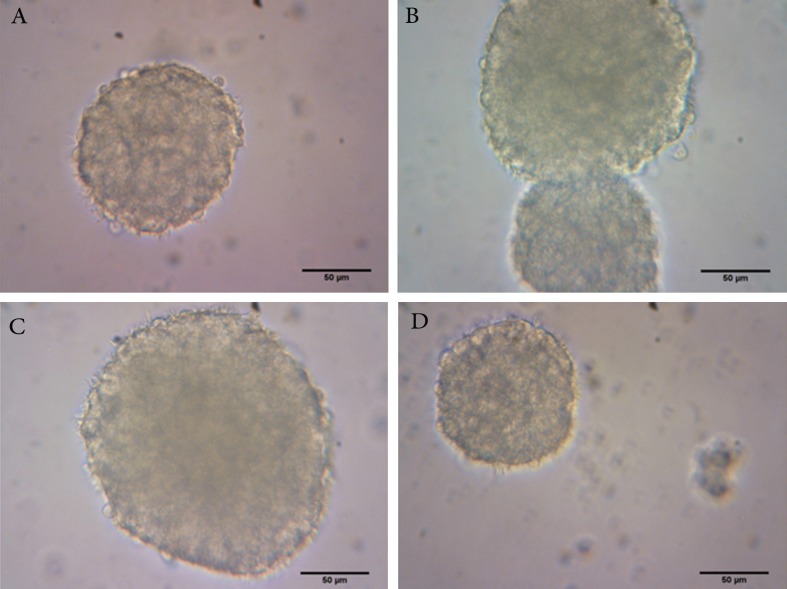
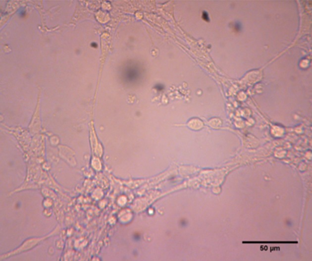
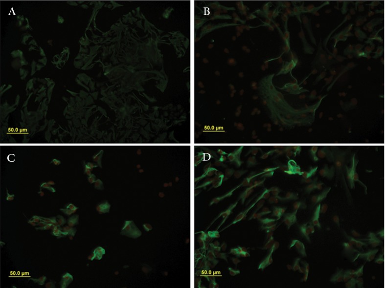
In this In this experimental study, we examined the role of e- CSF on proliferation and differentiation of neuroprogenitor cells using neurosphere culture method. Neurospheres size analysis and MTT assay were used to assess cell proliferation after four days in vitro. Glial differentiation study was carried out by immunocytochemistry. Neurospheres size and percentage of glial fibrialy acidic protein (GFAP) positive cells were measured by image analyzer (image J). The data were analyzed by one-way ANOVA, followed by the Tukey's post hoc test. Data were expressed as mean ± SEM, and differences were considered significant when p<0.05, 0.01 and 0.001.
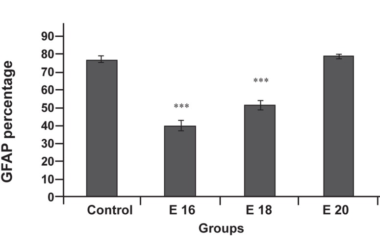
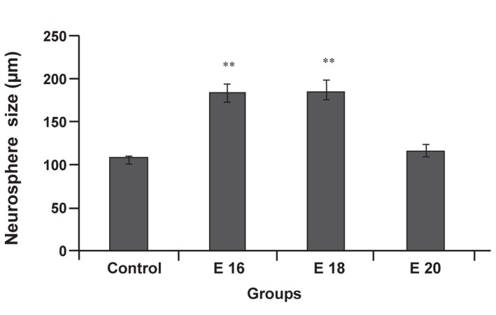
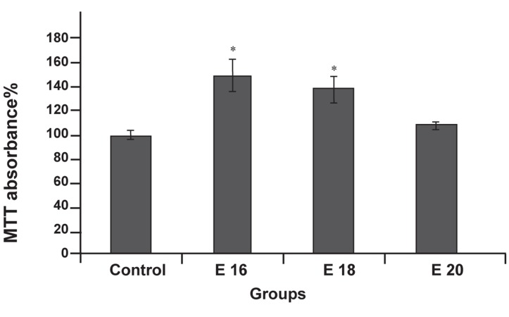
Viability and proliferation of neuro progenitor cells in cultures conditioned with E16 CSF and E18 CSF were significantly increased compare to control group. A dramatic decrease in percentage of GFAP-positive cells was found following the application of CSF from E16 and E18 embryos, but not E20 CSF.
Our data suggest that, e-CSF altered proliferation and differentiation of neuro progenitor cells in age dependent manner. E16 and E18 CSF enhanced proliferation and viability of neuro progenitor cells, and inhibited differentiation to glial fate in comparison with control group.
胚胎脑脊液(e-CSF)在胚胎和成人脑的发育中具有重要作用。蛋白质组学分析表明,这种液体中含有许多形态发生素和细胞因子,它们在胚胎生命的时间和空间中发生变化。本研究旨在评估胚胎 CSF 对不同胎龄神经祖细胞增殖和分化的发育影响。
在这项实验研究中,我们使用神经球培养法研究了 e-CSF 对神经祖细胞增殖和分化的作用。四体外培养后,通过神经球大小分析和 MTT 测定评估细胞增殖。通过免疫细胞化学研究神经球的胶质分化。通过图像分析仪(Image J)测量神经球大小和神经胶质纤维酸性蛋白(GFAP)阳性细胞的百分比。采用单因素方差分析,随后采用 Tukey 事后检验对数据进行分析。数据表示为均数±SEM,当 p<0.05、0.01 和 0.001 时,差异被认为具有统计学意义。
与对照组相比,培养物中 E16 CSF 和 E18 CSF 处理后的神经祖细胞活力和增殖明显增加。与 E16 和 E18 胚胎 CSF 相比,E20 CSF 应用后发现 GFAP 阳性细胞的百分比显著下降。
我们的数据表明,e-CSF 以年龄依赖的方式改变神经祖细胞的增殖和分化。与对照组相比,E16 和 E18 CSF 增强了神经祖细胞的增殖和活力,并抑制了向胶质命运的分化。