Nyholm Tufve, Jonsson Joakim, Söderström Karin, Bergström Per, Carlberg Andreas, Frykholm Gunilla, Behrens Claus F, Geertsen Poul Flemming, Trepiakas Redas, Hanvey Scott, Sadozye Azmat, Ansari Jawaher, McCallum Hazel, Frew John, McMenemin Rhona, Zackrisson Björn
Radiat Oncol. 2013 May 24;8:126. doi: 10.1186/1748-717X-8-126.
The use of magnetic resonance (MR) imaging as a part of preparation for radiotherapy is increasing. For delineation of the prostate several publications have shown decreased delineation variability using MR compared to computed tomography (CT). The purpose of the present work was to investigate the intra- and inter-physician delineation variability for prostate and seminal vesicles, and to investigate the influence of different MR sequence settings used clinically at the five centers participating in the study.
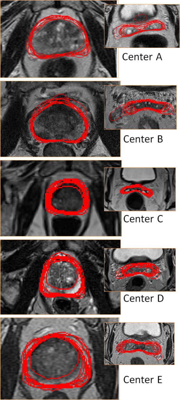
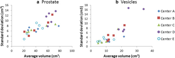
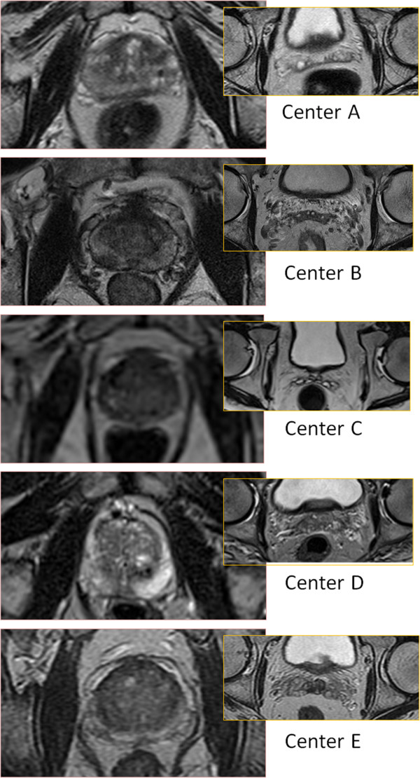
MR series from five centers, each providing five patients, were used. Two physicians from each center delineated the prostate and the seminal vesicles on each of the 25 image sets. The variability between the delineations was analyzed with respect to overall, intra- and inter-physician variability, and dependence between variability and origin of the MR images, i.e. the MR sequence used to acquire the data.
The intra-physician variability in different directions was between 1.3 - 1.9 mm and 3 - 4 mm for the prostate and seminal vesicles respectively (1 std). The inter-physician variability for different directions were between 0.7 - 1.7 mm and approximately equal for the prostate and seminal vesicles. Large differences in variability were observed for individual patients, and also for individual imaging sequences used at the different centers. There was however no indication of decreased variability with higher field strength.
The overall delineation variability is larger for the seminal vesicles compared to the prostate, due to a larger intra-physician variability. The imaging sequence appears to have a large influence on the variability, even for different variants of the T2-weighted spin-echo based sequences, which were used by all centers in the study.
磁共振(MR)成像作为放射治疗准备工作的一部分,其应用正在增加。对于前列腺的勾画,与计算机断层扫描(CT)相比,一些出版物表明使用MR可降低勾画的变异性。本研究的目的是调查医生内部和医生之间对前列腺和精囊的勾画变异性,并研究参与该研究的五个中心临床使用的不同MR序列设置的影响。
使用来自五个中心的MR系列,每个中心提供五名患者。每个中心的两名医生在25个图像集上分别勾画前列腺和精囊。分析了勾画之间的变异性,包括总体、医生内部和医生之间的变异性,以及变异性与MR图像来源(即用于获取数据的MR序列)之间的相关性。
对于前列腺和精囊,不同方向上医生内部的变异性分别在1.3 - 1.9毫米和3 - 4毫米之间(1个标准差)。不同方向上医生之间的变异性在0.7 - 1.7毫米之间,前列腺和精囊大致相等。在个体患者以及不同中心使用的个体成像序列中,观察到变异性存在很大差异。然而,没有迹象表明随着场强增加变异性会降低。
由于医生内部变异性较大,精囊的总体勾画变异性比前列腺大。成像序列似乎对变异性有很大影响,即使对于所有中心在研究中使用的基于T2加权自旋回波序列的不同变体也是如此。