Department of Anatomy and Neurobiology, Washington University School of Medicine St. Louis, MO, USA.
Front Neuroanat. 2013 May 14;7:7. doi: 10.3389/fnana.2013.00007. eCollection 2013.
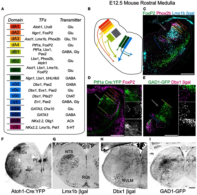
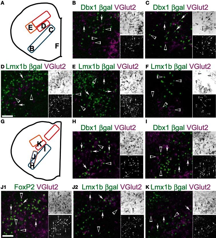
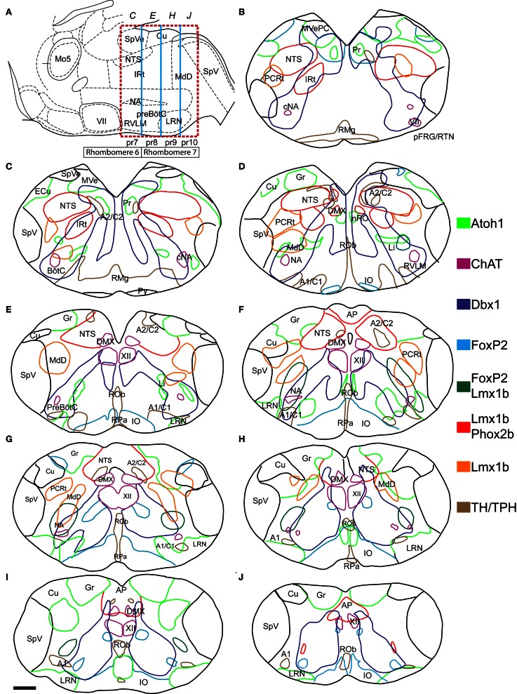
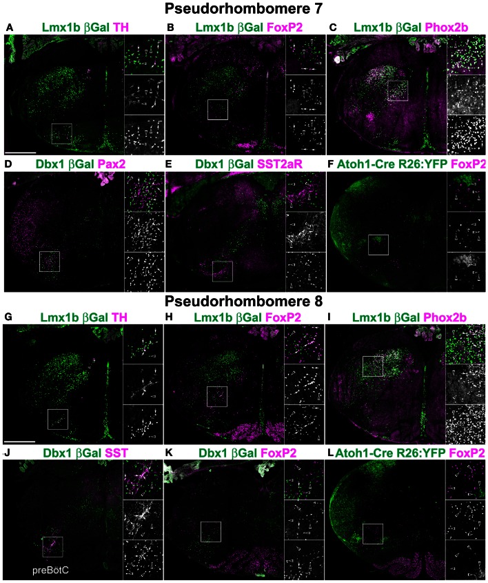
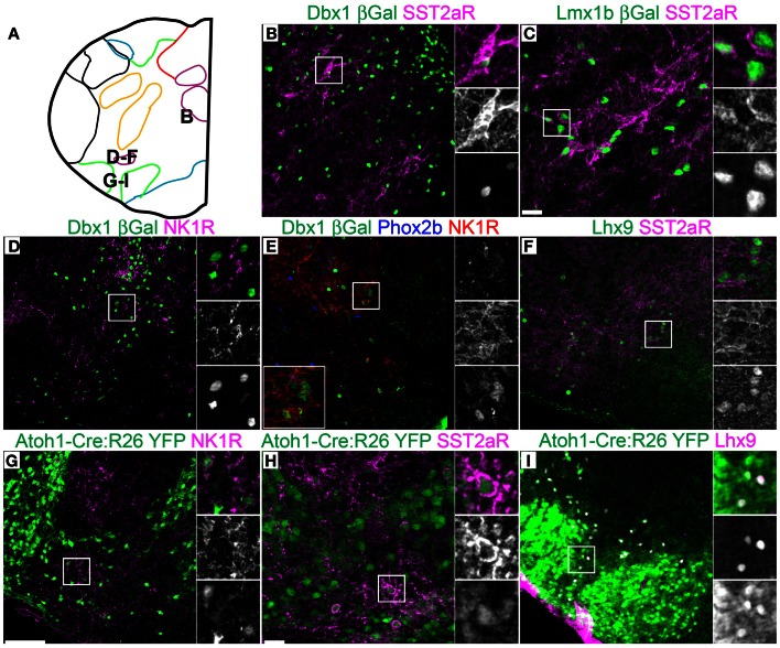
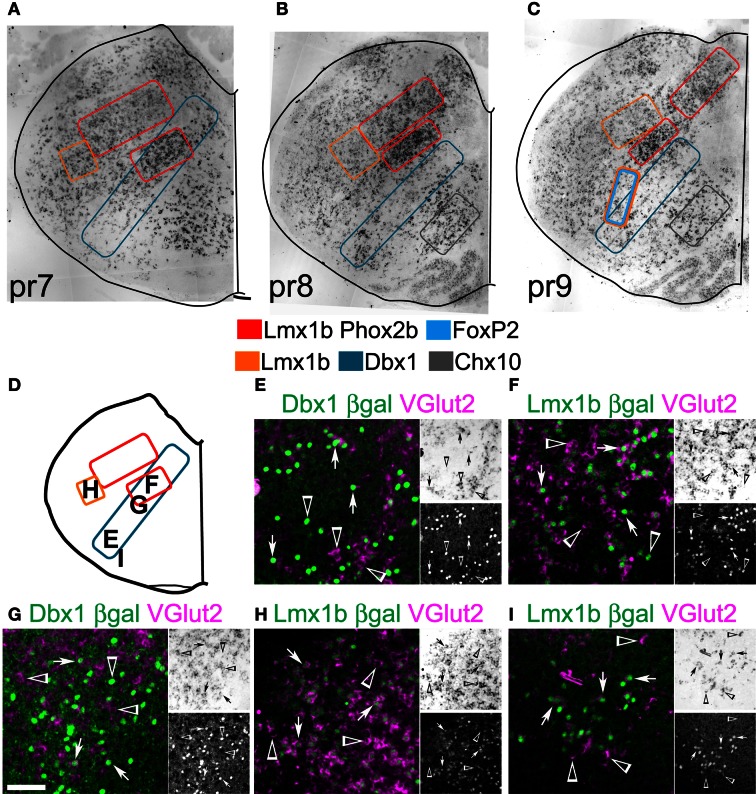
The medullary reticular formation contains large populations of inadequately described, excitatory interneurons that have been implicated in multiple homeostatic behaviors including breathing, viserosensory processing, vascular tone, and pain. Many hindbrain nuclei show a highly stereotyped pattern of localization across vertebrates suggesting a strong underlying genetic organization. Whether this is true for neurons within the reticular regions of hindbrain is unknown. Hindbrain neurons are derived from distinct developmental progenitor domains each of which expresses distinct patterns of transcription factors (TFs). These neuronal populations have distinct characteristics such as transmitter identity, migration, and connectivity suggesting developmentally expressed TFs might identify unique subpopulations of neurons within the reticular formation. A fate-mapping strategy using perinatal expression of reporter genes within Atoh1, Dbx1, Lmx1b, and Ptf1a transgenic mice coupled with immunohistochemistry (IHC) and in situ hybridization (ISH) were used to address the developmental organization of a large subset of reticular formation glutamatergic neurons. All hindbrain lineages have relatively large populations that extend the entire length of the hindbrain. Importantly, the location of neurons within each lineage was highly constrained. Lmx1b- and Dbx1- derived populations were both present in partially overlapping stripes within the reticular formation extending from dorsal to ventral brain. Within each lineage, distinct patterns of gene expression and organization were localized to specific hindbrain regions. Rostro-caudally sub-populations differ sequentially corresponding to proposed pseudo-rhombomereic boundaries. Dorsal-ventrally, sub-populations correspond to specific migratory positions. Together these data suggests the reticular formation is organized by a highly stereotyped developmental logic.
延髓网状结构包含大量描述不足的兴奋性中间神经元,这些神经元与多种稳态行为有关,包括呼吸、内脏感觉处理、血管张力和疼痛。许多后脑核在脊椎动物中表现出高度定型的定位模式,表明存在强烈的遗传组织。网状后脑区域内的神经元是否也是如此尚不清楚。后脑神经元来自不同的发育前体细胞区域,每个区域都表达不同的转录因子(TFs)模式。这些神经元群体具有不同的特征,如递质身份、迁移和连接性,表明发育表达的 TF 可能鉴定出网状结构内神经元的独特亚群。使用 Atoh1、Dbx1、Lmx1b 和 Ptf1a 转基因小鼠中的围产期报告基因表达进行的命运映射策略,结合免疫组织化学(IHC)和原位杂交(ISH),用于解决大的一部分网状形成谷氨酸能神经元的发育组织。所有后脑谱系都有相对较大的群体,延伸到整个后脑长度。重要的是,每个谱系内神经元的位置受到高度限制。Lmx1b 和 Dbx1 衍生的群体都存在于从背侧向腹侧脑延伸的网状结构中部分重叠的条纹内。在每个谱系中,基因表达和组织的独特模式都定位于特定的后脑区域。前后亚群依次对应于拟似菱形节边界。背腹侧,亚群对应于特定的迁移位置。这些数据表明,网状结构是由高度定型的发育逻辑组织的。