State Key Laboratory of Cognitive Neuroscience and Learning, Beijing Normal University, Beijing, China.
PLoS One. 2013 May 22;8(5):e63691. doi: 10.1371/journal.pone.0063691. Print 2013.
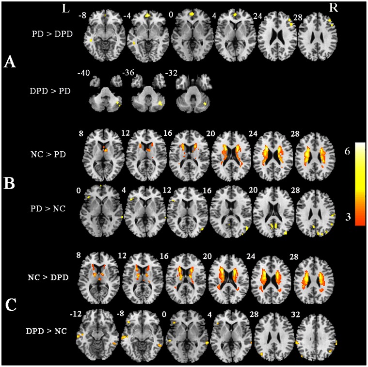
Depression is the most common psychiatric disorder observed in Parkinson's disease (PD) patients, however the neural contribution to the high rate of depression in the PD group is still unclear. In this study, we used resting-state functional magnetic resonance imaging (fMRI) to investigate the underlying neural mechanisms of depression in PD patients. Twenty-one healthy individuals and thirty-three patients with idiopathic PD, seventeen of whom were diagnosed with major depressive disorder, were recruited. An analysis of amplitude of low-frequency fluctuations (ALFF) was performed on the whole brain of all subjects. Our results showed that depressed PD patients had significantly decreased ALFF in the dorsolateral prefrontal cortex (DLPFC), the ventromedial prefrontal cortex (vMPFC) and the rostral anterior cingulated cortex (rACC) compared with non-depressed PD patients. A significant positive correlation was found between Hamilton Depression Rating Scale (HDRS) and ALFF in the DLPFC. The findings of changed ALFF in these brain regions implied depression in PD patients may be associated with abnormal activities of prefrontal-limbic network.
抑郁症是帕金森病(PD)患者中最常见的精神障碍,但 PD 患者中抑郁症高发的神经机制仍不清楚。在这项研究中,我们使用静息态功能磁共振成像(fMRI)来研究 PD 患者抑郁的潜在神经机制。我们招募了 21 名健康个体和 33 名特发性 PD 患者,其中 17 名被诊断为重度抑郁症。对所有受试者的全脑进行了低频振幅(ALFF)分析。结果显示,与非抑郁 PD 患者相比,抑郁 PD 患者的背外侧前额叶皮层(DLPFC)、腹内侧前额叶皮层(vMPFC)和前扣带皮层腹侧(rACC)的 ALFF 明显降低。汉密尔顿抑郁量表(HDRS)与 DLPFC 的 ALFF 呈显著正相关。这些脑区的 ALFF 变化表明 PD 患者的抑郁可能与前额叶-边缘网络的异常活动有关。