Department of Structural Biology, University of Pittsburgh School of Medicine, Pittsburgh, Pennsylvania 15260, USA.
Nature. 2013 May 30;497(7451):643-6. doi: 10.1038/nature12162.
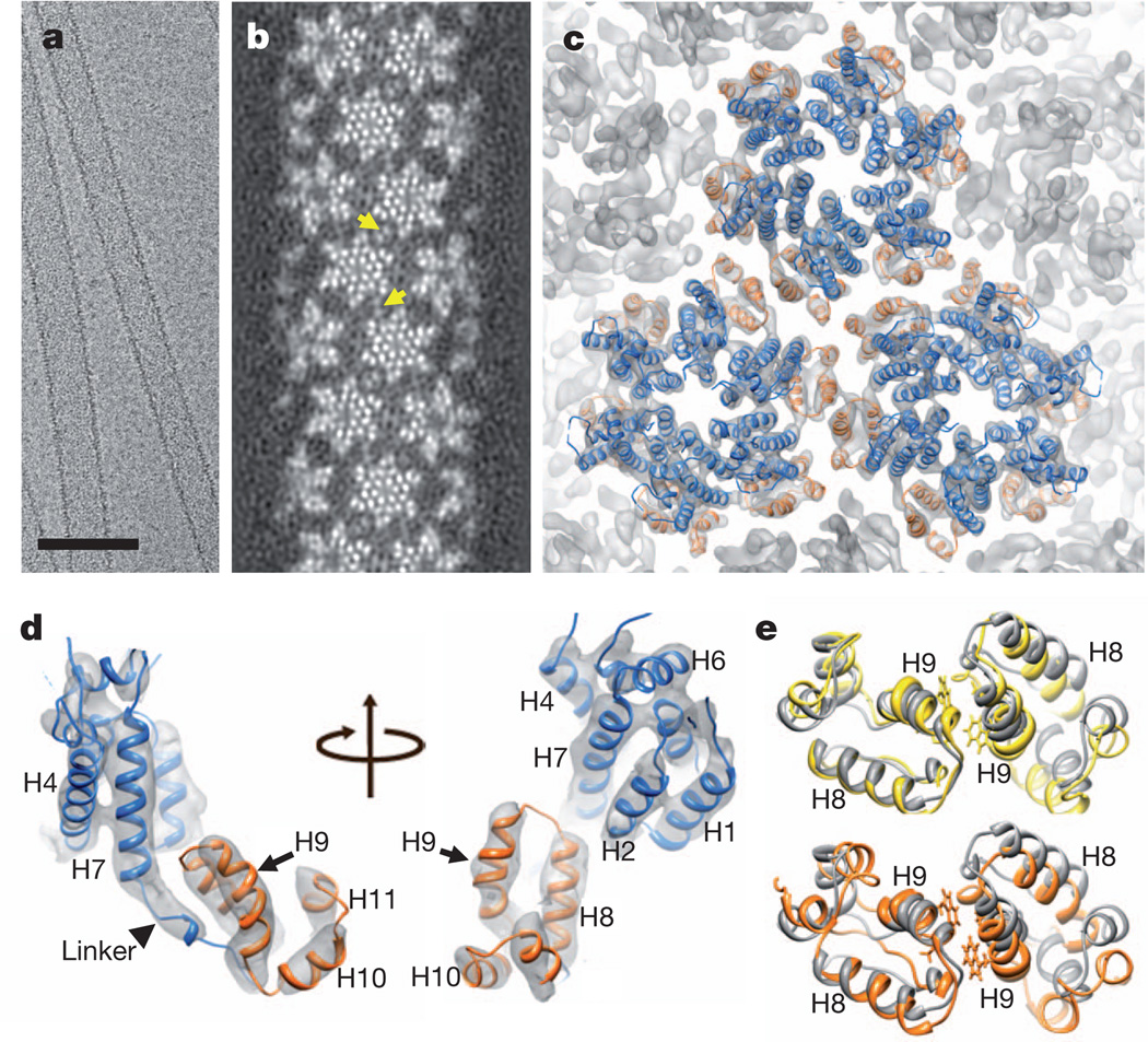
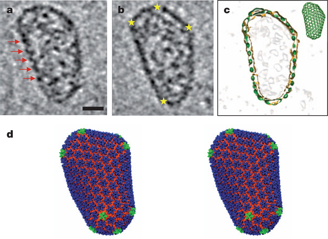
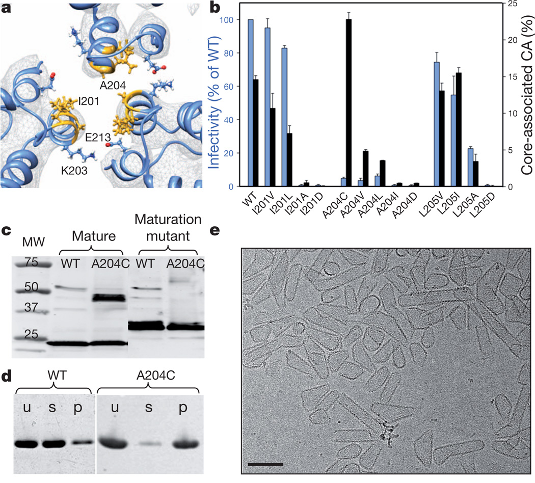
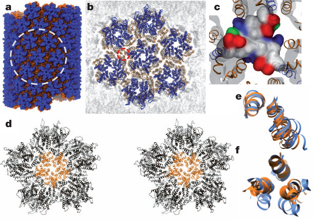
Retroviral capsid proteins are conserved structurally but assemble into different morphologies. The mature human immunodeficiency virus-1 (HIV-1) capsid is best described by a 'fullerene cone' model, in which hexamers of the capsid protein are linked to form a hexagonal surface lattice that is closed by incorporating 12 capsid-protein pentamers. HIV-1 capsid protein contains an amino-terminal domain (NTD) comprising seven α-helices and a β-hairpin, a carboxy-terminal domain (CTD) comprising four α-helices, and a flexible linker with a 310-helix connecting the two structural domains. Structures of the capsid-protein assembly units have been determined by X-ray crystallography; however, structural information regarding the assembled capsid and the contacts between the assembly units is incomplete. Here we report the cryo-electron microscopy structure of a tubular HIV-1 capsid-protein assembly at 8 Å resolution and the three-dimensional structure of a native HIV-1 core by cryo-electron tomography. The structure of the tubular assembly shows, at the three-fold interface, a three-helix bundle with critical hydrophobic interactions. Mutagenesis studies confirm that hydrophobic residues in the centre of the three-helix bundle are crucial for capsid assembly and stability, and for viral infectivity. The cryo-electron-microscopy structures enable modelling by large-scale molecular dynamics simulation, resulting in all-atom models for the hexamer-of-hexamer and pentamer-of-hexamer elements as well as for the entire capsid. Incorporation of pentamers results in closer trimer contacts and induces acute surface curvature. The complete atomic HIV-1 capsid model provides a platform for further studies of capsid function and for targeted pharmacological intervention.
逆转录病毒衣壳蛋白在结构上是保守的,但组装成不同的形态。成熟的人类免疫缺陷病毒 1(HIV-1)衣壳最适合用“富勒烯锥”模型来描述,其中衣壳蛋白的六聚体连接在一起,形成一个六方表面晶格,通过整合 12 个衣壳蛋白五聚体来封闭。HIV-1 衣壳蛋白包含一个由七个α-螺旋和一个β发夹组成的氨基末端结构域(NTD)、一个包含四个α-螺旋的羧基末端结构域(CTD),以及一个连接两个结构域的柔性连接体,其中有一个 310 螺旋。衣壳蛋白组装单元的结构已通过 X 射线晶体学确定;然而,关于组装衣壳和组装单元之间的接触的结构信息并不完整。在这里,我们报告了在 8Å分辨率下管状 HIV-1 衣壳蛋白组装的低温电子显微镜结构和通过低温电子断层扫描的天然 HIV-1 核心的三维结构。管状组装的结构在三折叠界面处显示出一个具有关键疏水相互作用的三螺旋束。突变研究证实,三螺旋束中心的疏水性残基对于衣壳组装和稳定性以及病毒感染力至关重要。低温电子显微镜结构使通过大规模分子动力学模拟进行建模成为可能,从而为六聚体-六聚体和五聚体-六聚体元件以及整个衣壳生成了全原子模型。五聚体的整合导致更紧密的三聚体接触并诱导急性表面曲率。完整的原子 HIV-1 衣壳模型为衣壳功能的进一步研究和靶向药物干预提供了一个平台。