• 文献检索
  • 文档翻译
  • 深度研究
  • 学术资讯
  • Suppr Zotero 插件Zotero 插件
  • 邀请有礼
  • 套餐&价格
  • 历史记录
应用&插件
Suppr Zotero 插件Zotero 插件浏览器插件Mac 客户端Windows 客户端微信小程序
定价
高级版会员购买积分包购买API积分包
服务
文献检索文档翻译深度研究API 文档MCP 服务
关于我们
关于 Suppr公司介绍联系我们用户协议隐私条款
关注我们

Suppr 超能文献

核心技术专利:CN118964589B侵权必究
粤ICP备2023148730 号-1Suppr @ 2026

文献检索

告别复杂PubMed语法,用中文像聊天一样搜索,搜遍4000万医学文献。AI智能推荐,让科研检索更轻松。

立即免费搜索

文件翻译

保留排版,准确专业,支持PDF/Word/PPT等文件格式,支持 12+语言互译。

免费翻译文档

深度研究

AI帮你快速写综述,25分钟生成高质量综述,智能提取关键信息,辅助科研写作。

立即免费体验

关节镜下切除外踝分离的小骨块。

Arthroscopic excision of separated ossicles of the lateral malleolus.

作者信息

Monden Satoshi, Hasegawa Atsushi, Hio Naohiro, Taki Masanori, Noguchi Hideo

机构信息

Kiryu Orthopedic Surgery Hospital, 284-1 Hirosawacho-Ainoshima, Kiryu, Gunma, 376-0014, Japan,

出版信息

J Orthop Sci. 2013 Sep;18(5):733-9. doi: 10.1007/s00776-013-0412-3. Epub 2013 Jun 1.

DOI:10.1007/s00776-013-0412-3
PMID:23728893
原文链接:https://pmc.ncbi.nlm.nih.gov/articles/PMC3778221/
Abstract

BACKGROUND

We have conducted a retrospective review of 19 patients for whom 20 separated ossicles of the lateral malleolus were excised arthroscopically. We examined the operating methods, findings, and overall results.

METHODS

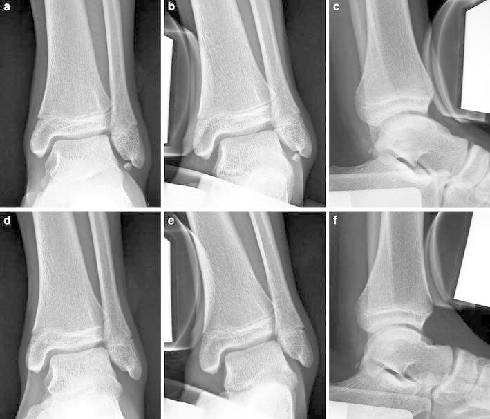
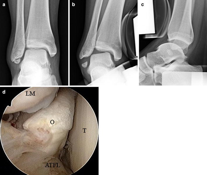
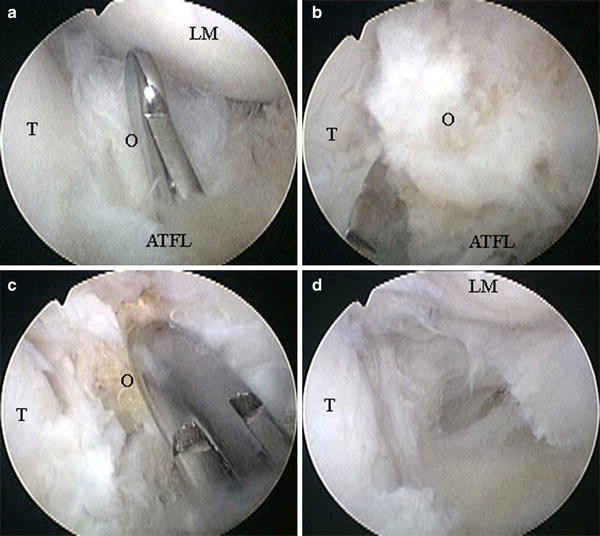
The patients' indications for this procedure were as follows. The main complaints were pain alone; ossicle sizes were small and ankle instability was minimal. There were 12 ankles of 12 males and eight ankles of seven females. The patients' average age was 17.6 years. A 2.7-mm, 30° arthroscope was inserted into the ankle joint through the anterolateral portal. Instruments were inserted through the accessory anterolateral portal, and ossicles were removed piece by piece. Talar tilt angles and anterior displacements were examined and compared before and after surgery by use of stress radiographs. Japanese Society for Surgery of the Foot (JSSF) ankle/hindfoot scales were assessed pre and postoperatively.

RESULTS

All patients recovered their original levels of activity. The mean talar tilt angle changed from 6.1° ± 2.4° preoperatively to 6.0° ± 1.8° postoperatively (p = 0.93), and the mean anterior displacement changed from 5.9 ± 1.7 mm preoperatively to 6.1 ± 2.0 mm postoperatively (p = 0.42). Average JSSF ankle/hindfoot scale improved from 77.6 ± 2.6 points preoperatively to 97.2 ± 5.2 points postoperatively (p < 0.01).

CONCLUSIONS

Arthroscopic excision of separated ossicles of the lateral malleolus achieved good results with minimum incisions, and relatively early resumption of daily and sports activity was possible. However, when the ossicles were embedded within the fibers of the anterior talofibular ligament, it was impossible to avoid cutting of ligament fibers. To reduce the possibility of ligament dysfunction, we believe postoperative treatment should conform to the accepted method for treatment of acute ankle sprains.

摘要

背景

我们对19例患者进行了回顾性研究,这些患者的20块分离的外踝小骨通过关节镜切除。我们检查了手术方法、发现结果及总体疗效。

方法

患者接受该手术的指征如下。主要症状仅为疼痛;小骨尺寸较小且踝关节不稳定程度轻微。其中男性12例共12个踝关节,女性7例共8个踝关节。患者平均年龄为17.6岁。通过前外侧入路将2.7毫米、30°的关节镜插入踝关节。器械通过辅助前外侧入路插入,小骨被逐块取出。术前和术后通过应力X线片检查并比较距骨倾斜角和前移情况。术前和术后评估日本足外科学会(JSSF)踝关节/后足评分标准。

结果

所有患者均恢复至原来的活动水平。平均距骨倾斜角术前为6.1°±2.4°,术后变为6.0°±1.8°(p = 0.93);平均前移术前为5.9±1.7毫米,术后变为6.1±2.0毫米(p = 0.42)。JSSF踝关节/后足评分标准平均得分术前为77.6±2.6分,术后提高至97.2±5.2分(p < 0.01)。

结论

关节镜下切除分离的外踝小骨切口最小,效果良好,且患者相对能够较早恢复日常活动和体育活动。然而,当小骨嵌入距腓前韧带纤维内时,无法避免切断韧带纤维。为降低韧带功能障碍的可能性,我们认为术后治疗应遵循公认的急性踝关节扭伤治疗方法。

https://cdn.ncbi.nlm.nih.gov/pmc/blobs/7161/3778221/0532e6bafb1e/776_2013_412_Fig3_HTML.jpg
https://cdn.ncbi.nlm.nih.gov/pmc/blobs/7161/3778221/1160aa2757ac/776_2013_412_Fig1_HTML.jpg
https://cdn.ncbi.nlm.nih.gov/pmc/blobs/7161/3778221/4c2444dec752/776_2013_412_Fig2_HTML.jpg
https://cdn.ncbi.nlm.nih.gov/pmc/blobs/7161/3778221/0532e6bafb1e/776_2013_412_Fig3_HTML.jpg
https://cdn.ncbi.nlm.nih.gov/pmc/blobs/7161/3778221/1160aa2757ac/776_2013_412_Fig1_HTML.jpg
https://cdn.ncbi.nlm.nih.gov/pmc/blobs/7161/3778221/4c2444dec752/776_2013_412_Fig2_HTML.jpg
https://cdn.ncbi.nlm.nih.gov/pmc/blobs/7161/3778221/0532e6bafb1e/776_2013_412_Fig3_HTML.jpg

相似文献

1
Arthroscopic excision of separated ossicles of the lateral malleolus.关节镜下切除外踝分离的小骨块。
J Orthop Sci. 2013 Sep;18(5):733-9. doi: 10.1007/s00776-013-0412-3. Epub 2013 Jun 1.
2
Arthroscopic Repair of Anterior Talofibular Ligament Using the Pull-Out Technique for Chronic Ankle Instability: Case Report.关节镜下采用拉出技术修复距腓前韧带治疗慢性踝关节不稳:病例报告
J Foot Ankle Surg. 2019 Jan;58(1):156-160. doi: 10.1053/j.jfas.2018.05.016. Epub 2018 Sep 20.
3
Comparison of the Modified Broström Procedure for Chronic Lateral Ankle Instability With and Without Subfibular Ossicle.伴有和不伴有腓骨下小骨的慢性外侧踝关节不稳改良 Broström 手术的比较
Am J Sports Med. 2016 Dec;44(12):3158-3164. doi: 10.1177/0363546516660069. Epub 2016 Aug 15.
4
The effect of an ossicle of the lateral malleolus on ligament reconstruction of chronic lateral ankle instability.外踝骨赘对慢性踝关节外侧不稳定韧带重建的影响。
Foot Ankle Int. 2010 Mar;31(3):191-6. doi: 10.3113/FAI.2010.0191.
5
A new minimally invasive method for anatomic reconstruction of the lateral ankle ligaments with a Tightrope system.一种使用Tightrope系统对踝关节外侧韧带进行解剖重建的新型微创方法。
Arch Orthop Trauma Surg. 2018 Nov;138(11):1549-1555. doi: 10.1007/s00402-018-2955-4. Epub 2018 Jun 6.
6
Safe angles of ATFL and CFL anchor insertion into anatomical attachment of fibula in a lateral ankle ligament repair.在外侧踝关节韧带修复中,距腓前韧带(ATFL)和跟腓韧带(CFL)锚钉插入腓骨解剖附着点的安全角度。
J Orthop Sci. 2021 Jan;26(1):156-161. doi: 10.1016/j.jos.2020.02.011. Epub 2020 Mar 20.
7
Comparison of All-Inside Arthroscopic and Open Techniques for the Modified Broström Procedure for Ankle Instability.全关节镜下与开放技术治疗踝关节不稳改良 Broström 手术的比较
Foot Ankle Int. 2016 Oct;37(10):1037-1045. doi: 10.1177/1071100716666508. Epub 2016 Sep 13.
8
Morphology of Anterior Talofibular Ligament After Arthroscopic Lateral Ankle Ligament Repair.关节镜下外侧踝关节韧带修复后距腓前韧带的形态学变化
Foot Ankle Int. 2020 Aug;41(8):993-1001. doi: 10.1177/1071100720920610. Epub 2020 Jun 8.
9
Investigation of Factors Affecting the Clinical Results of Arthroscopic Anterior Talofibular Ligament Repair for Chronic Lateral Ankle Instability.关节镜下距腓前韧带修复治疗慢性踝关节外侧不稳定的临床疗效影响因素研究。
J Foot Ankle Surg. 2020 May-Jun;59(3):465-468. doi: 10.1053/j.jfas.2019.09.010.
10
Prospective study of the " Inside-Out" arthroscopic ankle ligament technique: Preliminary result.“由内而外”关节镜下踝关节韧带技术的前瞻性研究:初步结果
Foot Ankle Surg. 2018 Aug;24(4):320-325. doi: 10.1016/j.fas.2017.03.002. Epub 2017 Mar 22.

引用本文的文献

1
Is it necessary to remove small fibular ossicles during an arthroscopic modified Broström operation for chronic lateral ankle instability?在关节镜下改良布罗斯特伦手术治疗慢性外侧踝关节不稳时,有必要切除小的腓骨小骨吗?
BMC Musculoskelet Disord. 2025 Apr 21;26(1):383. doi: 10.1186/s12891-025-08546-7.
2
Clinical outcomes over 2 years following arthroscopic ankle lateral ligament repair with os subfibulare.采用腓下骨块辅助关节镜下踝关节外侧韧带修复术后2年的临床疗效
Asia Pac J Sports Med Arthrosc Rehabil Technol. 2024 Nov 26;39:9-14. doi: 10.1016/j.asmart.2024.11.002. eCollection 2025 Jan.
3
Surgical treatment of ankle instability in children with os subfibulare.

本文引用的文献

1
The effect of an ossicle of the lateral malleolus on ligament reconstruction of chronic lateral ankle instability.外踝骨赘对慢性踝关节外侧不稳定韧带重建的影响。
Foot Ankle Int. 2010 Mar;31(3):191-6. doi: 10.3113/FAI.2010.0191.
2
Ossicles associated with chronic pain around the malleoli of the ankle.与踝关节周围慢性疼痛相关的小骨。
J Bone Joint Surg Br. 2008 Aug;90(8):1049-54. doi: 10.1302/0301-620X.90B8.20331.
3
Acute treatment of inversion ankle sprains: immobilization versus functional treatment.踝关节内翻扭伤的急性治疗:固定治疗与功能治疗对比
带副舟骨的儿童踝关节不稳定的手术治疗。
Arch Orthop Trauma Surg. 2023 Oct;143(10):6123-6129. doi: 10.1007/s00402-023-04905-y. Epub 2023 May 29.
4
Optimal Visualization of Os Subfibulare Using 3D Water Selective Cartilage Scan (3D_WATSc) MRI Sequencing: A Case Report.使用3D水选择性软骨扫描(3D_WATSc)MRI序列对腓下骨进行最佳可视化:一例报告
Cureus. 2022 Jul 29;14(7):e27469. doi: 10.7759/cureus.27469. eCollection 2022 Jul.
5
Surgical outcome of avulsion fractures of the distal fibula: a systematic literature review.腓骨远端撕脱性骨折的手术疗效:一项系统的文献综述
Arch Orthop Trauma Surg. 2023 Mar;143(3):1275-1291. doi: 10.1007/s00402-021-04256-6. Epub 2021 Nov 24.
6
Arthroscopic Stabilization of Unstable Os Subfibulare.关节镜下稳定不稳定的腓骨下籽骨
Arthrosc Tech. 2019 Sep 12;8(9):e1007-e1012. doi: 10.1016/j.eats.2019.05.011. eCollection 2019 Sep.
7
Anatomical variation in the ankle and foot: from incidental finding to inductor of pathology. Part I: ankle and hindfoot.踝关节和足部的解剖变异:从偶然发现到病理诱发因素。第一部分:踝关节和后足。
Insights Imaging. 2019 Jul 31;10(1):74. doi: 10.1186/s13244-019-0746-2.
8
Radiologic findings for prediction of rehabilitation outcomes in patients with chronic symptomatic os subfibulare.慢性症状性腓下籽骨患者康复结局预测的影像学表现
Radiol Med. 2017 Oct;122(10):766-773. doi: 10.1007/s11547-017-0786-y. Epub 2017 Jun 29.
9
Intraarticular Entrapment of Os Subfibulare Following a Severe Inversion Injury of the Ankle: A Case Report.踝关节严重内翻损伤后腓下籽骨关节内卡压:一例报告
Arch Trauma Res. 2015 Jun 20;4(2):e27046. doi: 10.5812/atr.4(2)2015.27046. eCollection 2015 Jun.
Clin Orthop Relat Res. 2007 Feb;455:169-72. doi: 10.1097/BLO.0b013e31802f5468.
4
A prospective, randomized clinical investigation of the treatment of first-time ankle sprains.首次踝关节扭伤治疗的前瞻性随机临床研究。
Am J Sports Med. 2006 Sep;34(9):1401-12. doi: 10.1177/0363546506288676. Epub 2006 Jun 26.
5
Evaluation of anterior talofibular ligament lesion using 3-dimensional computed tomography.使用三维计算机断层扫描评估距腓前韧带损伤
J Comput Assist Tomogr. 2006 May-Jun;30(3):543-7. doi: 10.1097/00004728-200605000-00032.
6
Development and reliability of a standard rating system for outcome measurement of foot and ankle disorders I: development of standard rating system.足踝疾病疗效评估标准评分系统的开发与可靠性 I:标准评分系统的开发
J Orthop Sci. 2005 Sep;10(5):457-65. doi: 10.1007/s00776-005-0936-2.
7
Symptomatic os subfibulare caused by accessory ossification: a case report.副骨化引起的有症状的腓下骨:一例报告
Clin Orthop Relat Res. 2002 Jun(399):197-200. doi: 10.1097/00003086-200206000-00023.
8
Treatment of ruptures of the lateral ankle ligaments: a meta-analysis.外侧踝关节韧带断裂的治疗:一项荟萃分析。
J Bone Joint Surg Am. 2000 Jun;82(6):761-73. doi: 10.2106/00004623-200006000-00002.
9
Arthroscopy of the ankle: analysis of results and indications on a series of 75 cases.
Foot Ankle Int. 1999 Nov;20(11):744-51. doi: 10.1177/107110079902001113.
10
Separated ossicles of the lateral malleolus.外踝的分离小骨。
Clin Orthop Relat Res. 1996 Sep(330):157-65. doi: 10.1097/00003086-199609000-00019.