Llanes Aaron Cedric D, Van Tassel Dane, Wirth Alexxa, Goncalves Luis F, Belthur Mohan V
Orthopaedic Surgery, University of Arizona College of Medicine-Phoenix, Phoenix, USA.
Radiology, Phoenix Children's Hospital, Phoenix, USA.
Cureus. 2022 Jul 29;14(7):e27469. doi: 10.7759/cureus.27469. eCollection 2022 Jul.
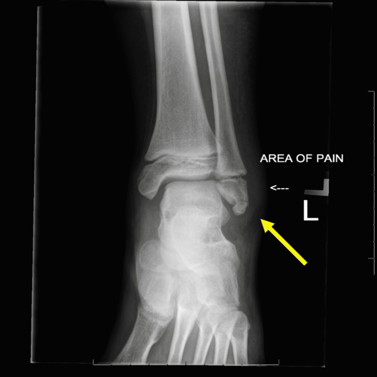
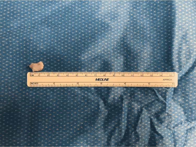
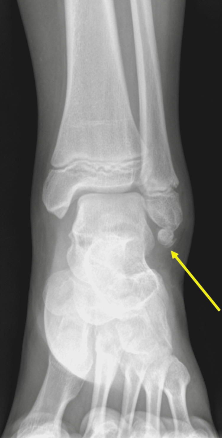
Os subfibulare is an accessory ossicle of the lateral malleolus at the distal end of the fibula. In most instances, os subfibulare is found incidentally on radiographs. While os subfibulare typically remains asymptomatic, some cases may present with ankle pain or instability. To initiate appropriate treatment and maximize patient outcomes, it is crucial to accurately visualize the accessory ossicle. Here, we report a symptomatic case of os subfibulare diagnosed with ankle radiographs and a 3D water selective cartilage scan (3D_WATSc, Ingenia, Philips Healthcare, The Netherlands) magnetic resonance imaging sequence and treated surgically with open ossicle excision and a modified Broström procedure.
腓下骨是位于腓骨远端外踝的一个附属小骨。在大多数情况下,腓下骨是在X线片上偶然发现的。虽然腓下骨通常无症状,但有些病例可能会出现踝关节疼痛或不稳定。为了启动适当的治疗并使患者获得最佳治疗效果,准确显示附属小骨至关重要。在此,我们报告一例经踝关节X线片和三维水选择性软骨扫描(3D_WATSc,荷兰飞利浦医疗保健公司Ingenia)磁共振成像序列诊断为腓下骨且有症状的病例,并通过开放性小骨切除术和改良的布罗斯特伦手术进行了手术治疗。