Translational Neuroscience Facility, School of Medical Sciences, University of New South Wales Sydney, NSW, Australia.
Front Neurol. 2013 May 20;4:58. doi: 10.3389/fneur.2013.00058. eCollection 2013.
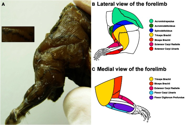
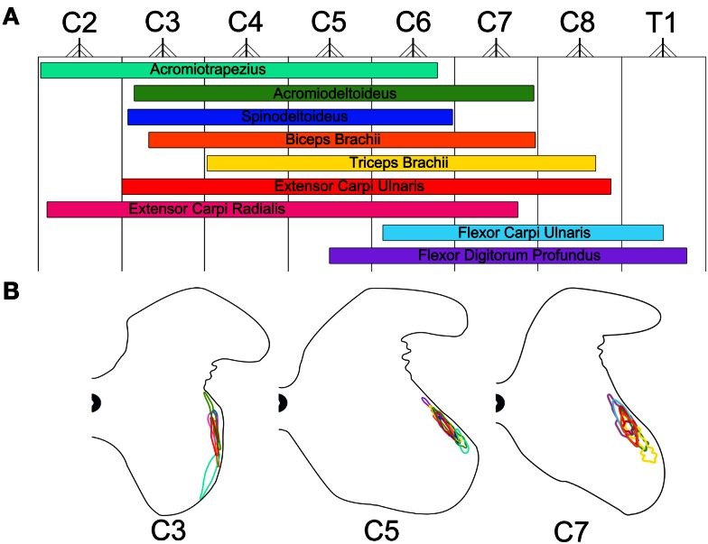
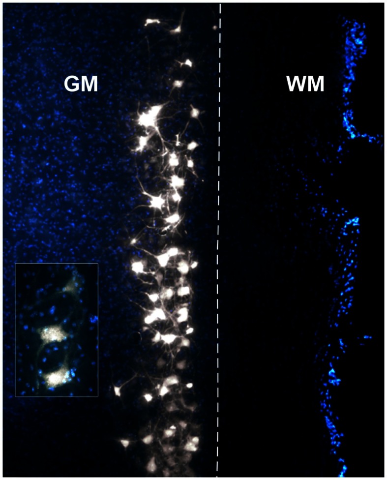
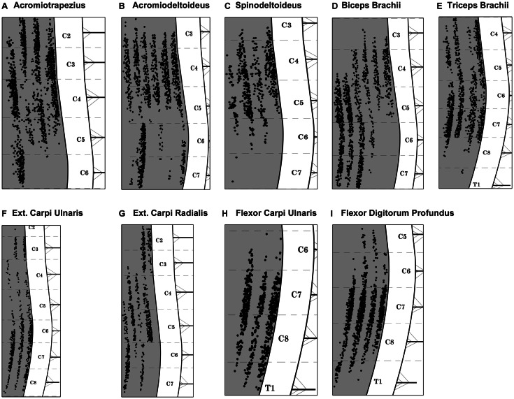
Lower motor neuron dysfunction is one of the most debilitating motor conditions. In this regard, transgenic mouse models of various lower motor neuron dysfunctions provide insight into the mechanisms underlying these pathologies and can also aid the development of new therapies. Viral-mediated gene therapy can take advantage of the muscle-motor neuron topographical relationship to shuttle therapeutic genes into specific populations of motor neurons in these mouse models. In this context, motor end plates (MEPs) are highly specialized regions on the skeletal musculature that offer direct access to the pre-synaptic nerve terminals, henceforth to the spinal cord motor neurons. The aim of this study was two-folded. First, it was to characterize the exact position of the MEP regions for several muscles of the mouse forelimb using acetylcholinesterase histochemistry. This MEP-muscle map was then used to guide a series of intramuscular injections of Fluoro-Gold (FG) in order to characterize the distribution of the innervating motor neurons. This analysis revealed that the MEPs are typically organized in an orthogonal fashion across the muscle fibers and extends throughout the full width of each muscle. Furthermore, targeting the full length of the MEP regions gave rise labeled motor neurons that are organized into columns spanning through more spinal cord segments than previously reported. The present analysis suggests that targeting the full width of the muscles' MEP regions with FG increases the somatic availability of the tracer. This process ensures a greater uptake of the tracer by the pre-synaptic nerve terminals, hence maximizing the labeling in spinal cord motor neurons. This investigation should have positive implications for future studies involving the somatic delivery of therapeutic genes into motor neurons for the treatment of various motor dysfunctions.
下运动神经元功能障碍是最具致残性的运动障碍之一。在这方面,各种下运动神经元功能障碍的转基因小鼠模型为这些病理的潜在机制提供了深入的了解,并且还可以帮助开发新的治疗方法。病毒介导的基因治疗可以利用肌肉-运动神经元的拓扑关系,将治疗基因穿梭到这些小鼠模型中特定的运动神经元群体中。在这种情况下,运动终板(MEP)是骨骼肌上高度特化的区域,可直接进入突触前神经末梢,进而进入脊髓运动神经元。本研究的目的有两个。首先,使用乙酰胆碱酯酶组织化学方法对小鼠前肢的几个肌肉的 MEP 区域的精确位置进行了描述。然后,将该 MEP-肌肉图谱用于指导一系列肌内注射荧光金(FG),以描述支配运动神经元的分布。该分析表明,MEP 通常以与肌肉纤维正交的方式组织,并延伸到每个肌肉的全宽。此外,靶向 MEP 区域的全长会导致标记的运动神经元呈柱状排列,跨越比以前报道的更多的脊髓节段。目前的分析表明,用 FG 靶向肌肉 MEP 区域的全长会增加示踪剂的躯体可用性。该过程确保了更多的示踪剂被突触前神经末梢摄取,从而使脊髓运动神经元的标记最大化。这项研究应该对未来涉及将治疗基因通过躯体递送到运动神经元以治疗各种运动障碍的研究具有积极意义。