Department of Pathology, University of New Mexico Albuquerque, NM, USA.
Front Oncol. 2013 May 17;3:97. doi: 10.3389/fonc.2013.00097. eCollection 2013.
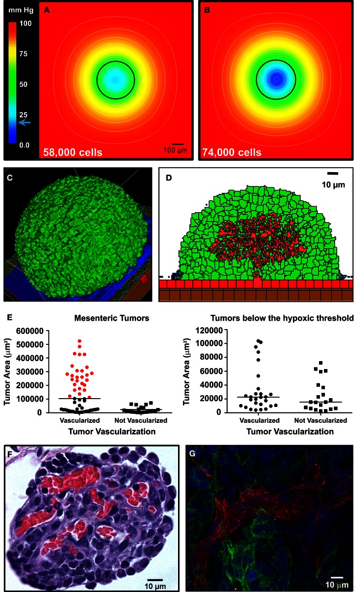
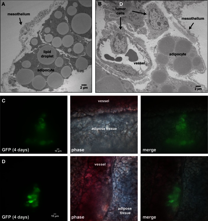
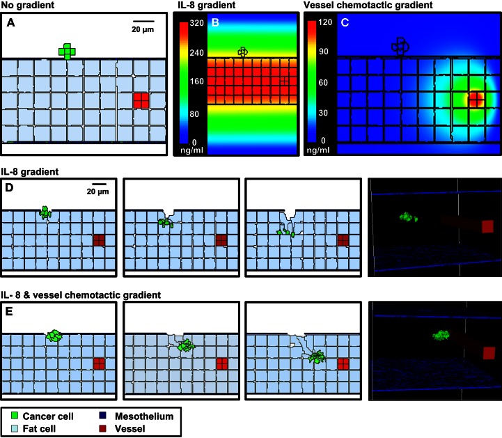
Ovarian cancer relapse is often characterized by metastatic spread throughout the peritoneal cavity with tumors attached to multiple organs. In this study, interaction of ovarian cancer cells with the peritoneal tumor microenvironment was evaluated in a xenograft model based on intraperitoneal injection of fluorescent SKOV3.ip1 ovarian cancer cells. Intra-vital microscopy of mixed GFP-red fluorescent protein (RFP) cell populations injected into the peritoneum demonstrated that cancer cells aggregate and attach as mixed spheroids, emphasizing the importance of homotypic adhesion in tumor formation. Electron microscopy provided high resolution structural information about local attachment sites. Experimental measurements from the mouse model were used to build a three-dimensional cellular Potts ovarian tumor model (OvTM) that examines ovarian cancer cell attachment, chemotaxis, growth, and vascularization. OvTM simulations provide insight into the relative influence of cancer cell-cell adhesion, oxygen availability, and local architecture on tumor growth and morphology. Notably, tumors on the mesentery, omentum, or spleen readily invade the "open" architecture, while tumors attached to the gut encounter barriers that restrict invasion and instead rapidly expand into the peritoneal space. Simulations suggest that rapid neovascularization of SKOV3.ip1 tumors is triggered by constitutive release of angiogenic factors in the absence of hypoxia. This research highlights the importance of cellular adhesion and tumor microenvironment in the seeding of secondary ovarian tumors on diverse organs within the peritoneal cavity. Results of the OvTM simulations indicate that invasion is strongly influenced by features underlying the mesothelial lining at different sites, but is also affected by local production of chemotactic factors. The integrated in vivo mouse model and computer simulations provide a unique platform for evaluating targeted therapies for ovarian cancer relapse.
卵巢癌复发的特征通常是肿瘤附着在多个器官上,通过腹腔转移扩散。在这项研究中,通过向腹腔内注射荧光 SKOV3.ip1 卵巢癌细胞,在基于腹腔内注射的异种移植模型中评估了卵巢癌细胞与腹腔肿瘤微环境的相互作用。对注射到腹膜中的混合 GFP-红色荧光蛋白(RFP)细胞群进行活体显微镜检查,结果表明癌细胞聚集并附着为混合球体,这强调了同种型粘附在肿瘤形成中的重要性。电子显微镜提供了有关局部附着部位的高分辨率结构信息。从小鼠模型获得的实验测量结果用于构建一个三维细胞 Potts 卵巢肿瘤模型(OvTM),该模型检查卵巢癌细胞附着、趋化性、生长和血管生成。OvTM 模拟提供了对癌细胞-细胞粘附、氧气可用性和局部结构对肿瘤生长和形态的相对影响的深入了解。值得注意的是,肠系膜、大网膜或脾脏上的肿瘤很容易侵入“开放”结构,而附着在肠道上的肿瘤则遇到限制其侵入的障碍,反而迅速扩展到腹腔空间。模拟表明,在缺氧的情况下,SKOV3.ip1 肿瘤中血管生成因子的持续释放会触发快速的血管生成。这项研究强调了细胞粘附和肿瘤微环境在卵巢癌复发时在不同器官上播散继发性卵巢肿瘤的重要性。OvTM 模拟的结果表明,侵袭强烈受到不同部位间皮衬里下潜在特征的影响,但也受到局部趋化因子产生的影响。体内小鼠模型和计算机模拟的综合提供了一个独特的平台,可用于评估针对卵巢癌复发的靶向治疗。