• 文献检索
  • 文档翻译
  • 深度研究
  • 学术资讯
  • Suppr Zotero 插件Zotero 插件
  • 邀请有礼
  • 套餐&价格
  • 历史记录
应用&插件
Suppr Zotero 插件Zotero 插件浏览器插件Mac 客户端Windows 客户端微信小程序
定价
高级版会员购买积分包购买API积分包
服务
文献检索文档翻译深度研究API 文档MCP 服务
关于我们
关于 Suppr公司介绍联系我们用户协议隐私条款
关注我们

Suppr 超能文献

核心技术专利:CN118964589B侵权必究
粤ICP备2023148730 号-1Suppr @ 2026

文献检索

告别复杂PubMed语法,用中文像聊天一样搜索,搜遍4000万医学文献。AI智能推荐,让科研检索更轻松。

立即免费搜索

文件翻译

保留排版,准确专业,支持PDF/Word/PPT等文件格式,支持 12+语言互译。

免费翻译文档

深度研究

AI帮你快速写综述,25分钟生成高质量综述,智能提取关键信息,辅助科研写作。

立即免费体验

基于患者校准的导管原位癌(DCIS)的代理建模:从微观测量到临床进展的宏观预测。

Patient-calibrated agent-based modelling of ductal carcinoma in situ (DCIS): from microscopic measurements to macroscopic predictions of clinical progression.

机构信息

Center for Applied Molecular Medicine, Keck School of Medicine, University of Southern California, Los Angeles, CA, USA.

出版信息

J Theor Biol. 2012 May 21;301:122-40. doi: 10.1016/j.jtbi.2012.02.002. Epub 2012 Feb 9.

DOI:10.1016/j.jtbi.2012.02.002
PMID:22342935
原文链接:https://pmc.ncbi.nlm.nih.gov/articles/PMC3322268/
Abstract

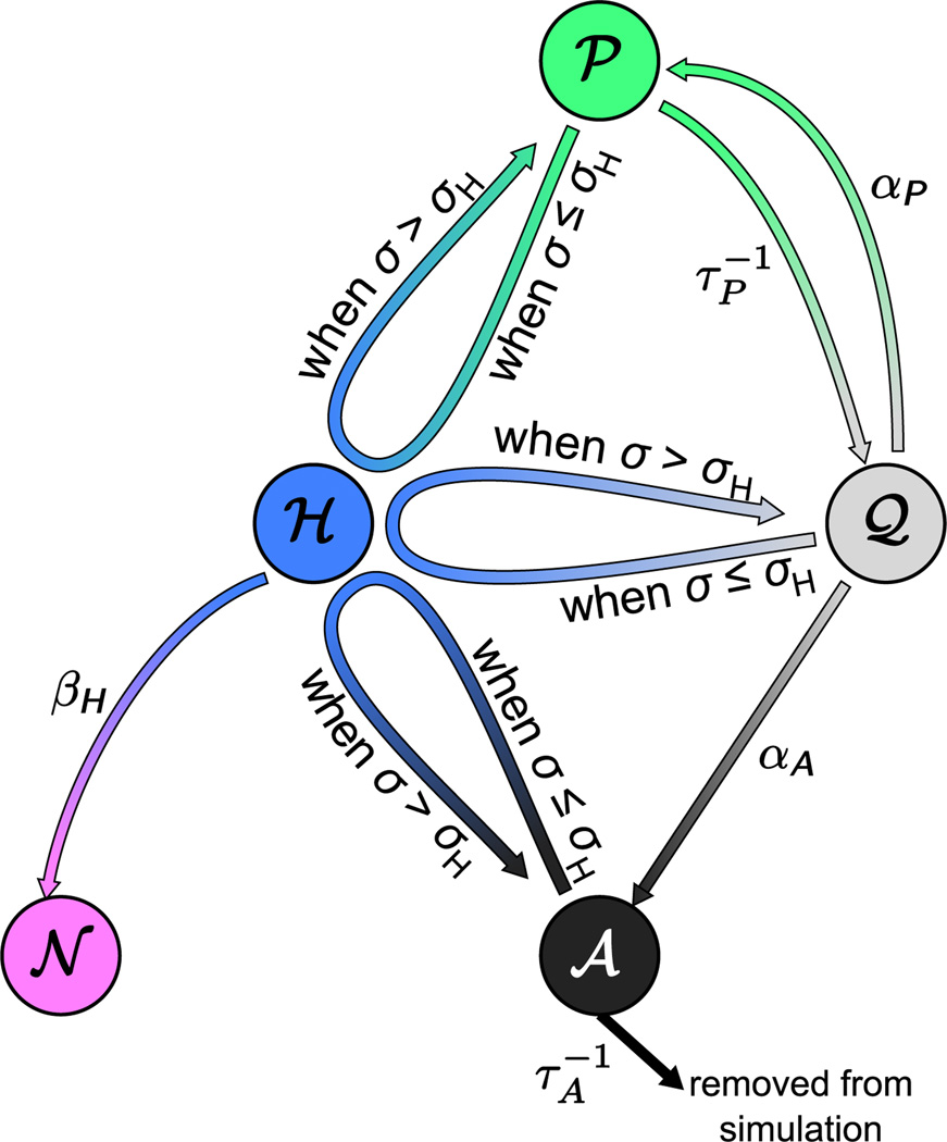
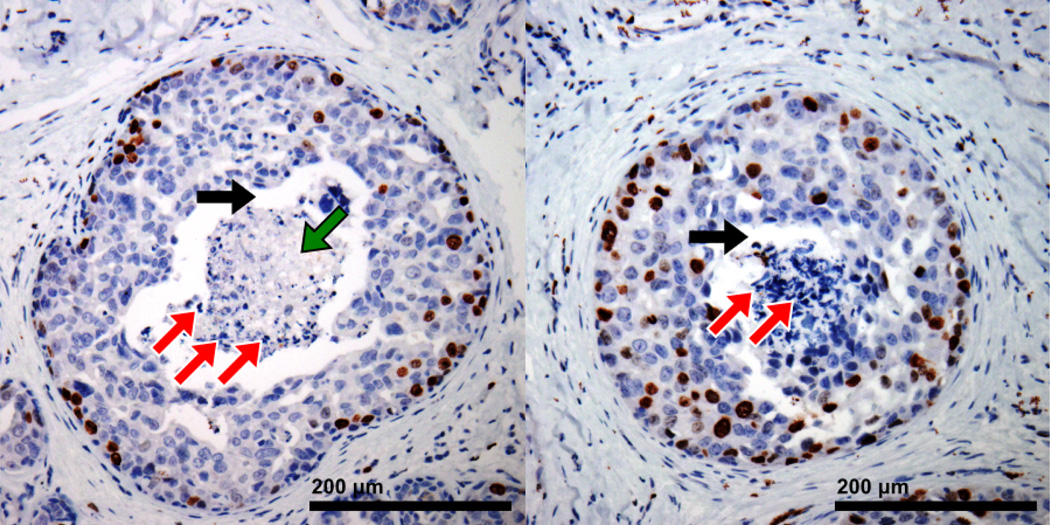
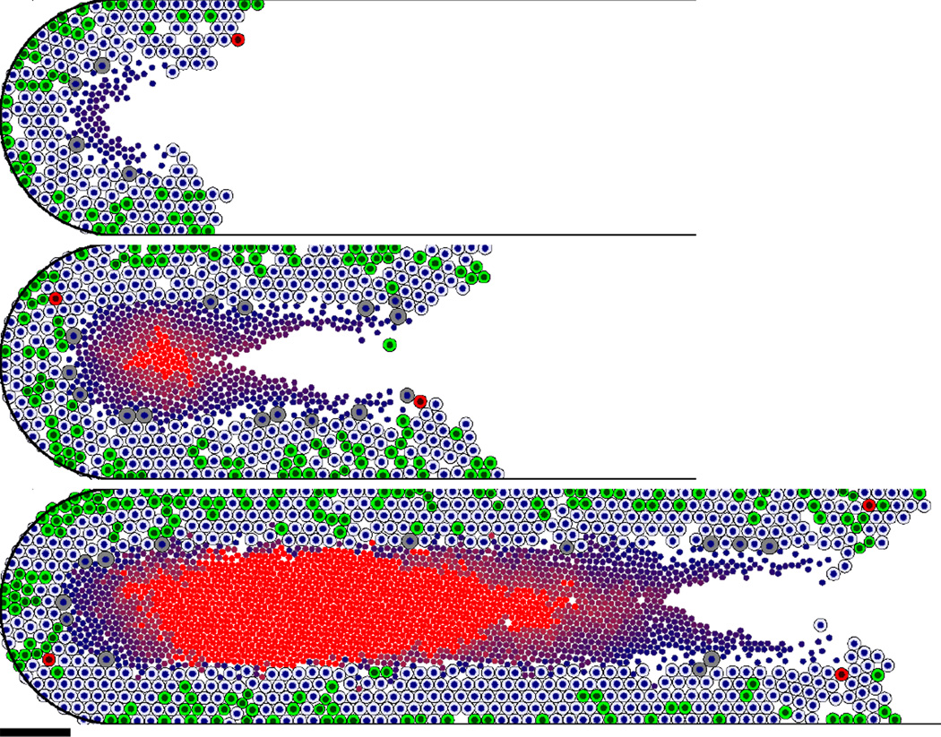
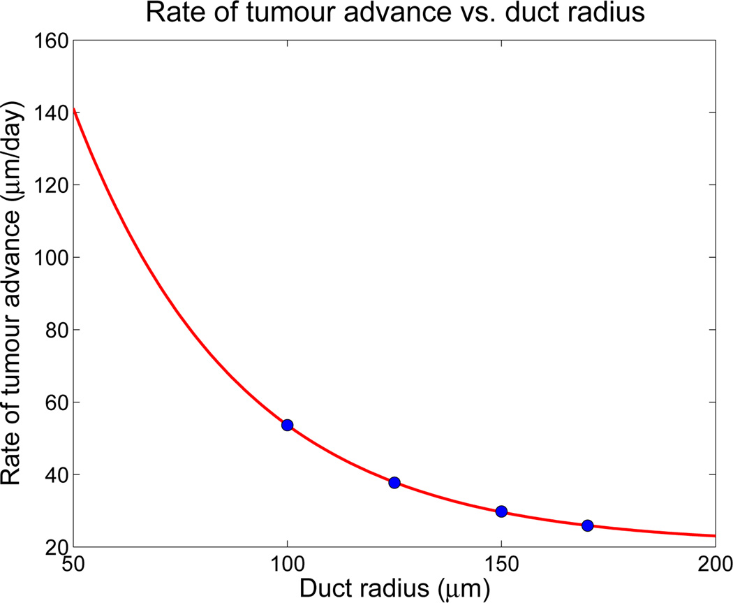
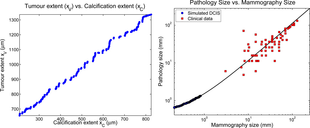
Ductal carcinoma in situ (DCIS)--a significant precursor to invasive breast cancer--is typically diagnosed as microcalcifications in mammograms. However, the effective use of mammograms and other patient data to plan treatment has been restricted by our limited understanding of DCIS growth and calcification. We develop a mechanistic, agent-based cell model and apply it to DCIS. Cell motion is determined by a balance of biomechanical forces. We use potential functions to model interactions with the basement membrane and amongst cells of unequal size and phenotype. Each cell's phenotype is determined by genomic/proteomic- and microenvironment-dependent stochastic processes. Detailed "sub-models" describe cell volume changes during proliferation and necrosis; we are the first to account for cell calcification. We introduce the first patient-specific calibration method to fully constrain the model based upon clinically-accessible histopathology data. After simulating 45 days of solid-type DCIS with comedonecrosis, the model predicts: necrotic cell lysis acts as a biomechanical stress relief and is responsible for the linear DCIS growth observed in mammography; the rate of DCIS advance varies with the duct radius; the tumour grows 7-10mm per year--consistent with mammographic data; and the mammographic and (post-operative) pathologic sizes are linearly correlated--in quantitative agreement with the clinical literature. Patient histopathology matches the predicted DCIS microstructure: an outer proliferative rim surrounds a stratified necrotic core with nuclear debris on its outer edge and calcification in the centre. This work illustrates that computational modelling can provide new insight on the biophysical underpinnings of cancer. It may 1-day be possible to augment a patient's mammography and other imaging with rigorously-calibrated models that help select optimal surgical margins based upon the patient's histopathologic data.

摘要

导管原位癌(DCIS)——一种显著的浸润性乳腺癌前体——通常在乳房 X 光片中被诊断为微钙化。然而,由于我们对 DCIS 生长和钙化的理解有限,乳房 X 光片和其他患者数据的有效利用一直受到限制。我们开发了一种基于机制的、基于代理的细胞模型,并将其应用于 DCIS。细胞运动由生物力学力的平衡决定。我们使用势函数来模拟与基底膜的相互作用以及大小和表型不等的细胞之间的相互作用。每个细胞的表型由基因组/蛋白质组和微环境依赖的随机过程决定。详细的“子模型”描述了细胞在增殖和坏死过程中的体积变化;我们是第一个考虑细胞钙化的。我们引入了第一个基于患者的校准方法,该方法基于临床可获得的组织病理学数据来完全约束模型。在模拟具有粉刺样坏死的实体型 DCIS 45 天后,该模型预测:坏死细胞裂解作为生物力学的应激缓解,负责在乳房 X 光片中观察到的线性 DCIS 生长;DCIS 进展的速度随导管半径而变化;肿瘤每年生长 7-10mm——与乳房 X 光数据一致;并且乳房 X 光和(术后)病理大小呈线性相关——与临床文献定量一致。患者组织病理学与预测的 DCIS 微观结构相匹配:增生性边缘环绕着分层坏死核心,其外边缘有核碎片,中心有钙化。这项工作表明,计算建模可以为癌症的生物物理基础提供新的见解。有朝一日,可能可以通过严格校准的模型来增强患者的乳房 X 光和其他成像,根据患者的组织病理学数据帮助选择最佳的手术边界。

https://cdn.ncbi.nlm.nih.gov/pmc/blobs/1c2f/3322268/ce3c9e630cdd/nihms356621f14.jpg
https://cdn.ncbi.nlm.nih.gov/pmc/blobs/1c2f/3322268/fe66cfd8eff1/nihms356621f1.jpg
https://cdn.ncbi.nlm.nih.gov/pmc/blobs/1c2f/3322268/540525911b64/nihms356621f2.jpg
https://cdn.ncbi.nlm.nih.gov/pmc/blobs/1c2f/3322268/c2ab1541f5d6/nihms356621f3.jpg
https://cdn.ncbi.nlm.nih.gov/pmc/blobs/1c2f/3322268/d37bb9e39e9a/nihms356621f4.jpg
https://cdn.ncbi.nlm.nih.gov/pmc/blobs/1c2f/3322268/29d636bb307e/nihms356621f5.jpg
https://cdn.ncbi.nlm.nih.gov/pmc/blobs/1c2f/3322268/bcb85c4ca0e5/nihms356621f6.jpg
https://cdn.ncbi.nlm.nih.gov/pmc/blobs/1c2f/3322268/15fef472dbed/nihms356621f7.jpg
https://cdn.ncbi.nlm.nih.gov/pmc/blobs/1c2f/3322268/f2962b9097e0/nihms356621f8.jpg
https://cdn.ncbi.nlm.nih.gov/pmc/blobs/1c2f/3322268/714c0beca08d/nihms356621f9.jpg
https://cdn.ncbi.nlm.nih.gov/pmc/blobs/1c2f/3322268/7f45ad8fcbd2/nihms356621f10.jpg
https://cdn.ncbi.nlm.nih.gov/pmc/blobs/1c2f/3322268/8d53e8b421d4/nihms356621f11.jpg
https://cdn.ncbi.nlm.nih.gov/pmc/blobs/1c2f/3322268/09a9dbd78ed9/nihms356621f12.jpg
https://cdn.ncbi.nlm.nih.gov/pmc/blobs/1c2f/3322268/974892a89f6f/nihms356621f13.jpg
https://cdn.ncbi.nlm.nih.gov/pmc/blobs/1c2f/3322268/ce3c9e630cdd/nihms356621f14.jpg

相似文献

1
Patient-calibrated agent-based modelling of ductal carcinoma in situ (DCIS): from microscopic measurements to macroscopic predictions of clinical progression.基于患者校准的导管原位癌(DCIS)的代理建模:从微观测量到临床进展的宏观预测。
J Theor Biol. 2012 May 21;301:122-40. doi: 10.1016/j.jtbi.2012.02.002. Epub 2012 Feb 9.
2
Microcalcifications associated with ductal carcinoma in situ: mammographic-pathologic correlation.与导管原位癌相关的微钙化:乳腺钼靶-病理相关性
Semin Diagn Pathol. 1994 Aug;11(3):181-92.
3
Patterns of aggressiveness: risk of progression to invasive breast cancer by mammographic features of calcifications in screen-detected ductal carcinoma in situ.形态学侵袭性:乳腺 X 线筛查下原位导管癌的钙化形态特征与浸润性乳腺癌进展风险的关系。
Acta Radiol. 2022 May;63(5):586-595. doi: 10.1177/02841851211006319. Epub 2021 Apr 22.
4
Sensitivity of breast MRI for ductal carcinoma in situ appearing as microcalcifications only on mammography.乳腺磁共振成像(MRI)对仅在乳腺钼靶上表现为微钙化的导管原位癌的敏感性。
Clin Imaging. 2016 Nov-Dec;40(6):1207-1212. doi: 10.1016/j.clinimag.2016.08.001. Epub 2016 Aug 6.
5
Qualitative Radiogenomics: Association Between BI-RADS Calcification Descriptors and Recurrence Risk as Assessed by the Oncotype DX Ductal Carcinoma In Situ Score.定性放射组学:BI-RADS 钙化描述符与 Oncotype DX 导管原位癌评分评估的复发风险之间的关联。
AJR Am J Roentgenol. 2019 Apr;212(4):919-924. doi: 10.2214/AJR.18.20306. Epub 2019 Feb 4.
6
Understanding Ductal Carcinoma In Situ Invasion using a Multiscale Agent-Based Model.使用基于多尺度智能体模型理解导管原位癌侵袭
Annu Int Conf IEEE Eng Med Biol Soc. 2018 Jul;2018:5846-5849. doi: 10.1109/EMBC.2018.8513542.
7
Mammographic extent of microcalcifications and oestrogen receptor expression affect preoperative breast carcinoma in situ size estimation.乳腺钼靶检查中微钙化的范围和雌激素受体表达影响术前原位乳腺癌大小的估计。
Breast Cancer. 2017 May;24(3):466-472. doi: 10.1007/s12282-016-0729-9. Epub 2016 Sep 17.
8
Lesion size is a major determinant of the mammographic features of ductal carcinoma in situ: findings from the Sloane project.病灶大小是导管原位癌乳腺 X 线特征的主要决定因素:Sloane 项目的研究结果。
Clin Radiol. 2010 Mar;65(3):181-4. doi: 10.1016/j.crad.2009.05.017. Epub 2010 Jan 18.
9
Can Occult Invasive Disease in Ductal Carcinoma In Situ Be Predicted Using Computer-extracted Mammographic Features?能否使用计算机提取的乳房X线摄影特征来预测导管原位癌中的隐匿性浸润性疾病?
Acad Radiol. 2017 Sep;24(9):1139-1147. doi: 10.1016/j.acra.2017.03.013. Epub 2017 May 11.
10
A mass on breast imaging predicts coexisting invasive carcinoma in patients with a core biopsy diagnosis of ductal carcinoma in situ.乳腺影像上的肿物可预测在粗针活检诊断为导管原位癌的患者中并存浸润性癌。
Am Surg. 2001 Sep;67(9):907-12.

引用本文的文献

1
A hybrid computational model of cancer spheroid growth with ribose-induced collagen stiffening.一种具有核糖诱导胶原蛋白硬化的癌症球体生长混合计算模型。
Front Bioeng Biotechnol. 2025 Apr 9;13:1515962. doi: 10.3389/fbioe.2025.1515962. eCollection 2025.
2
Breast cancer extracellular matrix invasion depends on local mechanical loading of the collagen network.乳腺癌细胞对细胞外基质的侵袭取决于胶原网络的局部机械负荷。
J Mater Chem B. 2025 Jul 10;13(27):8068-8082. doi: 10.1039/d4tb01474j.
3
Validating the predictions of mathematical models describing tumor growth and treatment response.

本文引用的文献

1
A novel, patient-specific mathematical pathology approach for assessment of surgical volume: application to ductal carcinoma in situ of the breast.一种新颖的、针对患者的数学病理学方法,用于评估手术量:在乳腺导管原位癌中的应用。
Anal Cell Pathol (Amst). 2011;34(5):247-63. doi: 10.3233/ACP-2011-0019.
2
The role of the microenvironment in tumor growth and invasion.微环境在肿瘤生长和侵袭中的作用。
Prog Biophys Mol Biol. 2011 Aug;106(2):353-79. doi: 10.1016/j.pbiomolbio.2011.06.006. Epub 2011 Jun 28.
3
Multiscale cancer modeling.多尺度癌症建模。
验证描述肿瘤生长和治疗反应的数学模型的预测结果。
ArXiv. 2025 Feb 26:arXiv:2502.19333v1.
4
Biopsy location and tumor-associated macrophages in predicting malignant glioma recurrence using an in-silico model.使用计算机模拟模型,活检部位和肿瘤相关巨噬细胞在预测恶性胶质瘤复发中的作用
NPJ Syst Biol Appl. 2025 Jan 8;11(1):3. doi: 10.1038/s41540-024-00478-7.
5
A multiscale model of immune surveillance in micrometastases gives insights on cancer patient digital twins.微转移中免疫监视的多尺度模型为癌症患者数字孪生提供了见解。
NPJ Syst Biol Appl. 2024 Dec 4;10(1):144. doi: 10.1038/s41540-024-00472-z.
6
Agent-Based Modeling of Virtual Tumors Reveals the Critical Influence of Microenvironmental Complexity on Immunotherapy Efficacy.基于主体的虚拟肿瘤建模揭示了微环境复杂性对免疫治疗疗效的关键影响。
Cancers (Basel). 2024 Aug 23;16(17):2942. doi: 10.3390/cancers16172942.
7
A comprehensive review of computational cell cycle models in guiding cancer treatment strategies.计算细胞周期模型在指导癌症治疗策略中的综合综述。
NPJ Syst Biol Appl. 2024 Jul 5;10(1):71. doi: 10.1038/s41540-024-00397-7.
8
In silico study of heterogeneous tumour-derived organoid response to CAR T-cell therapy.基于计算机的异质性肿瘤类器官对 CAR T 细胞治疗反应的研究。
Sci Rep. 2024 May 29;14(1):12307. doi: 10.1038/s41598-024-63125-5.
9
A seven-step guide to spatial, agent-based modelling of tumour evolution.肿瘤演化的基于主体的空间建模七步指南。
Evol Appl. 2024 May 2;17(5):e13687. doi: 10.1111/eva.13687. eCollection 2024 May.
10
Agent-based modeling in cancer biomedicine: applications and tools for calibration and validation.基于代理的癌症生物医学建模:校准和验证的应用和工具。
Cancer Biol Ther. 2024 Dec 31;25(1):2344600. doi: 10.1080/15384047.2024.2344600. Epub 2024 Apr 28.
Annu Rev Biomed Eng. 2011 Aug 15;13:127-55. doi: 10.1146/annurev-bioeng-071910-124729.
4
Mammographic morphology and distribution of calcifications in ductal carcinoma in situ diagnosed in organized screening.在有组织的筛查中诊断出的导管原位癌的乳腺钼靶形态及钙化分布
Acta Radiol. 2011 Jun 1;52(5):481-7. doi: 10.1258/ar.2011.100357. Epub 2011 Mar 28.
5
A New Ghost Cell/Level Set Method for Moving Boundary Problems: Application to Tumor Growth.一种用于移动边界问题的新型幽灵单元/水平集方法:在肿瘤生长中的应用。
J Sci Comput. 2008 Jun 1;35(2-3):266-299. doi: 10.1007/s10915-008-9190-z.
6
Re-resection rates after breast-conserving surgery as a performance indicator: introduction of a case-mix model to allow comparison between Dutch hospitals.保乳手术后再次切除率作为绩效指标:引入病例组合模型以允许荷兰各医院之间进行比较。
Eur J Surg Oncol. 2011 Apr;37(4):357-63. doi: 10.1016/j.ejso.2011.01.008. Epub 2011 Feb 2.
7
Nonlinear modelling of cancer: bridging the gap between cells and tumours.癌症的非线性建模:弥合细胞与肿瘤之间的差距。
Nonlinearity. 2010;23(1):R1-R9. doi: 10.1088/0951-7715/23/1/r01.
8
A theoretical quantitative model for evolution of cancer chemotherapy resistance.癌症化疗耐药性演变的理论定量模型。
Biol Direct. 2010 Apr 20;5:25. doi: 10.1186/1745-6150-5-25.
9
Lesion size is a major determinant of the mammographic features of ductal carcinoma in situ: findings from the Sloane project.病灶大小是导管原位癌乳腺 X 线特征的主要决定因素:Sloane 项目的研究结果。
Clin Radiol. 2010 Mar;65(3):181-4. doi: 10.1016/j.crad.2009.05.017. Epub 2010 Jan 18.
10
A 2D mechanistic model of breast ductal carcinoma in situ (DCIS) morphology and progression.二维乳腺导管原位癌(DCIS)形态和进展的机理模型。
J Theor Biol. 2010 Apr 21;263(4):393-406. doi: 10.1016/j.jtbi.2009.11.024. Epub 2009 Dec 16.