Singh Ashok, Singh Anupama, Sand Jordan M, Heninger Erika, Hafeez Bilal Bin, Verma Ajit K
Department of Human Oncology, Wisconsin Institutes for Medical Research, School of Medicine and Public Health, 1111 Highland Avenue, University of Wisconsin, Madison, WI 53705, USA.
J Skin Cancer. 2013;2013:452425. doi: 10.1155/2013/452425. Epub 2013 Apr 29.
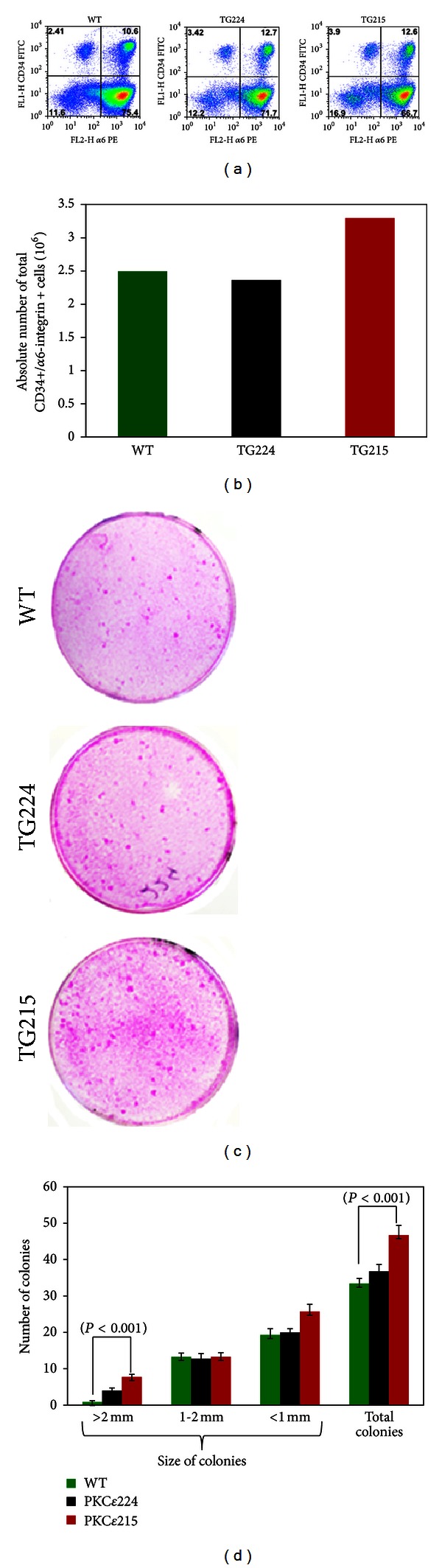
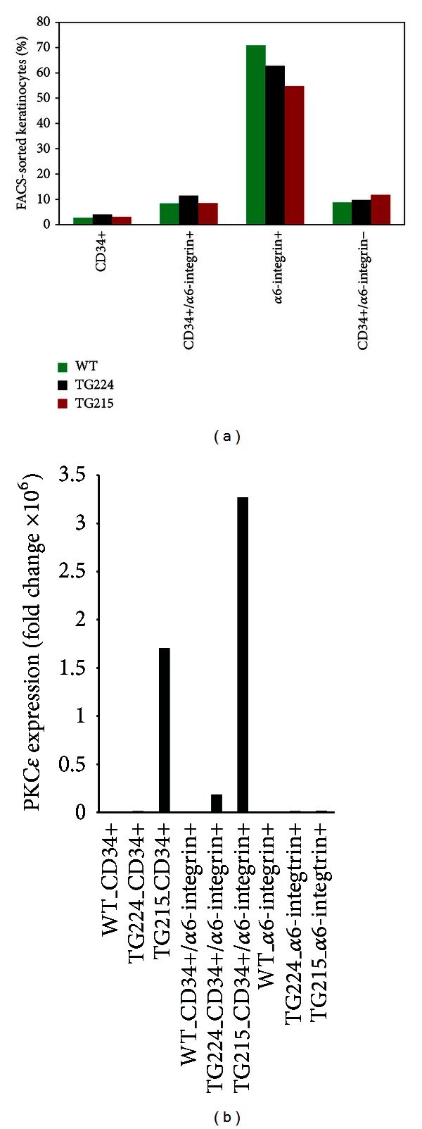
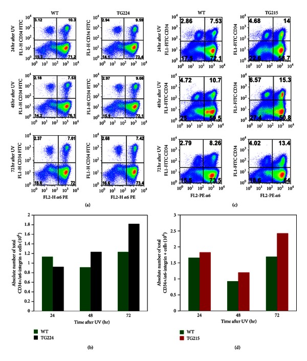
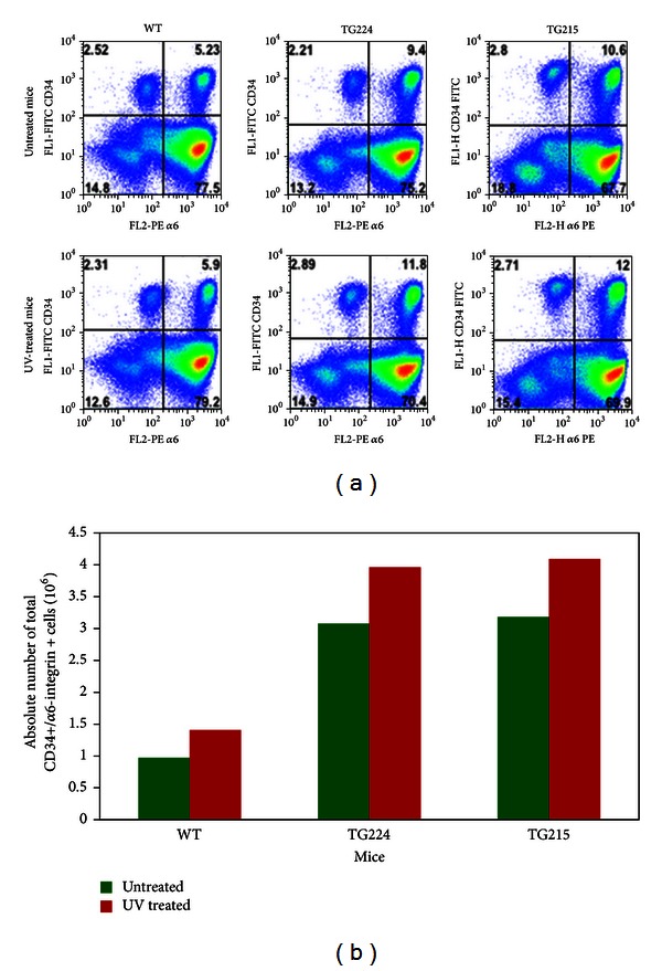
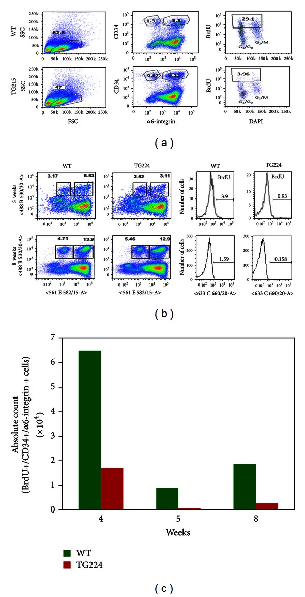
To find clues about the mechanism by which kinase C epsilon (PKC ε ) may impart susceptibility to ultraviolet radiation (UVR)-induced development of cutaneous squamous cell carcinomas (SCC), we compared PKC ε transgenic (TG) mice and their wild-type (WT) littermates for (1) the effects of UVR exposures on percent of putative hair follicle stem cells (HSCs) and (2) HSCs proliferation. The percent of double HSCs (CD34+ and α 6-integrin or CD34+/CD49f+) in the isolated keratinocytes were determined by flow cytometric analysis. Both single and chronic UVR treatments (1.8 kJ/m(2)) resulted in an increase in the frequency of double positive HSCs in PKC ε TG mice as compared to their WT littermates. To determine the rate of proliferation of bulge region stem cells, a 5-bromo-2'-deoxyuridine labeling (BrdU) experiment was performed. In the WT mice, the percent of double positive HSCs retaining BrdU label was 28.4 ± 0.6% compared to 4.0 ± 0.06% for the TG mice, an approximately 7-fold decrease. A comparison of gene expression profiles of FACS sorted double positive HSCs showed increased expression of Pes1, Rad21, Tfdp1 and Cks1b genes in TG mice compared to WT mice. Also, PKC ε over expression in mice increased the clonogenicity of isolated keratinocytes, a property commonly ascribed to stem cells.
为了找到蛋白激酶Cε(PKCε)可能赋予皮肤鳞状细胞癌(SCC)对紫外线辐射(UVR)诱导发展易感性的机制线索,我们比较了PKCε转基因(TG)小鼠及其野生型(WT)同窝小鼠在以下两方面的情况:(1)UVR照射对假定毛囊干细胞(HSCs)百分比的影响;(2)HSCs的增殖。通过流式细胞术分析确定分离的角质形成细胞中双阳性HSCs(CD34 +和α6整合素或CD34 + / CD49f +)的百分比。与WT同窝小鼠相比,单次和慢性UVR处理(1.8 kJ/m²)均导致PKCε TG小鼠中双阳性HSCs频率增加。为了确定隆突区干细胞的增殖速率,进行了5-溴-2'-脱氧尿苷标记(BrdU)实验。在WT小鼠中,保留BrdU标记的双阳性HSCs百分比为28.4±0.6%,而TG小鼠为4.0±0.06%,下降了约7倍。对通过荧光激活细胞分选术(FACS)分选的双阳性HSCs的基因表达谱进行比较,结果显示与WT小鼠相比,TG小鼠中Pes1、Rad21、Tfdp1和Cks1b基因的表达增加。此外,小鼠中PKCε的过表达增加了分离的角质形成细胞的克隆形成能力,这是干细胞通常具有的特性。