Pattern Recognition Laboratory, FAU Erlangen-Nuremberg, Martensstrasse 3, Erlangen 91058, Germany.
EJNMMI Res. 2013 Jun 5;3(1):45. doi: 10.1186/2191-219X-3-45.
Routine single-photon emission computed tomography (SPECT) currently lacks quantitative information on regional activity concentration (ACC) of the injected tracer (e.g. kBq/ml). Furthermore, little is known on the skeletal absolute concentration of 99mTc-DPD after intravenous injection in bone scintigraphy. The aim of this study is to determine ACC in the healthy lumbar vertebrae of patients using a recently published quantitative SPECT/computed tomography (CT) protocol.
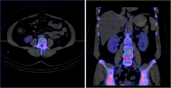
Lumbar vertebrae ACC estimates were performed in 50 female patients (mean age 69.88 ± 13.73 years) who had been administered 562.84 ± 102.33 MBq of 99mTc-DPD and had undergone SPECT acquisition 4 h after the injection. The SPECT/CT system was calibrated against a well counter. Images were reconstructed with Flash3D. ACC and CT tissue density were measured in volumes of interest drawn over the spongious bone tissue of the three lower lumbar vertebral bodies when these exhibited no focal CT or SPECT pathology.
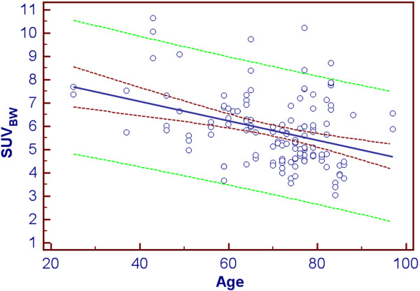
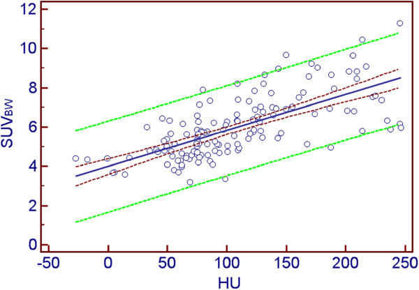
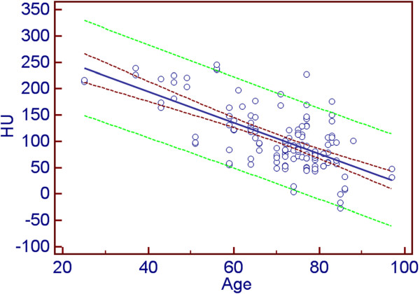
Average ACC measured in the normal spongious bone tissue was 48.15 ± 13.66 kBq/ml (95% confidence interval (CI) 45.81 to 50.50 kBq/ml). This corresponds to a mean standardised uptake value (SUV) of (5.91 ± 1.54) (95% CI (5.64 to 6.17) SUV). SUV correlated significantly with Hounsfield units (HU) (r = 0.678, p < 0.0001). Significant negative correlations were observed between age and HU (r = -0.650, p < 0.0001) and between age and SUV (r = -0.385, p < 0.0001).
The SUVs determined for 99mTc-DPD uptake 4 h post injection are in the same range as those reported for [18F]fluoride in positron emission tomography. The strong correlation of SUV with bone CT density underlines the physiological significance of this variable. Our data suggest further investigation of the potential value of ACC measurement in the diagnosis of pathological conditions such as osteoporosis or in following up osseous metastases under therapy.
目前,常规单光子发射计算机断层扫描(SPECT)缺乏关于注射示踪剂(例如 kBq/ml)的局部活性浓度(ACC)的定量信息。此外,静脉注射 99mTc-DPD 后在骨闪烁照相术中骨骼的绝对浓度知之甚少。本研究的目的是使用最近发表的定量 SPECT/计算机断层扫描(CT)方案来确定健康腰椎的 ACC。
对 50 名女性患者(平均年龄 69.88 ± 13.73 岁)进行腰椎 ACC 估计,这些患者已注射 562.84 ± 102.33 MBq 99mTc-DPD,并在注射后 4 小时进行 SPECT 采集。SPECT/CT 系统经过计数器校准。使用 Flash3D 进行图像重建。在没有焦点 CT 或 SPECT 病理的情况下,当三个下腰椎椎体的海绵骨组织显示无异常时,通过感兴趣区域(ROI)测量 ACC 和 CT 组织密度。
正常海绵骨组织中的平均 ACC 测量值为 48.15 ± 13.66 kBq/ml(95%置信区间(CI)为 45.81 至 50.50 kBq/ml)。这相当于平均标准化摄取值(SUV)为(5.91 ± 1.54)(95%CI(5.64 至 6.17)SUV)。SUV 与 Hounsfield 单位(HU)显著相关(r=0.678,p<0.0001)。年龄与 HU(r=-0.650,p<0.0001)和年龄与 SUV(r=-0.385,p<0.0001)之间存在显著负相关。
注射后 4 小时测定的 99mTc-DPD 摄取 SUV 值与正电子发射断层扫描中报告的 [18F] 氟化物值处于相同范围。SUV 与骨 CT 密度的强烈相关性强调了该变量的生理意义。我们的数据表明,进一步研究 ACC 测量在诊断骨质疏松症等病理状况或在治疗后监测骨转移方面的潜在价值。