Department of Fragrance and Cosmetic Science, Kaohsiung Medical University, Kaohsiung, Taiwan.
PLoS One. 2013 Jun 10;8(6):e56330. doi: 10.1371/journal.pone.0056330. Print 2013.
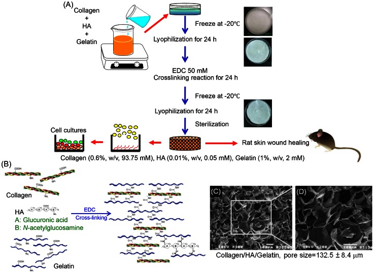
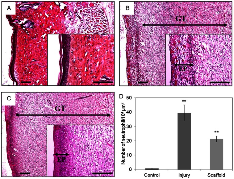
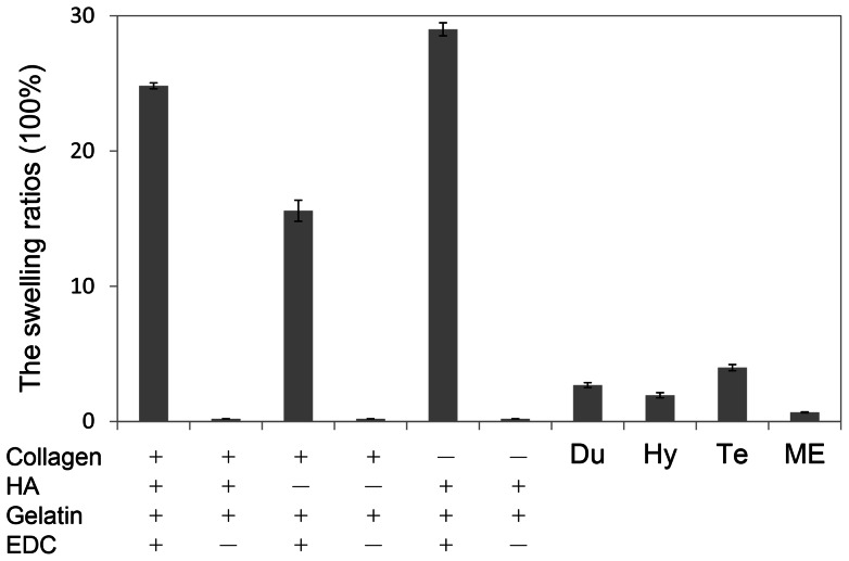
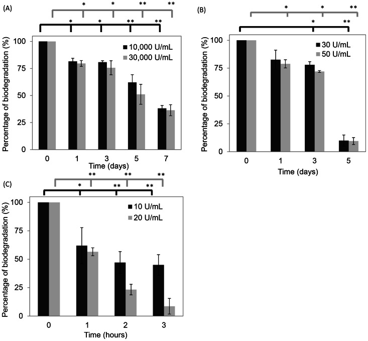
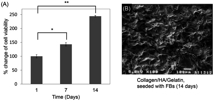
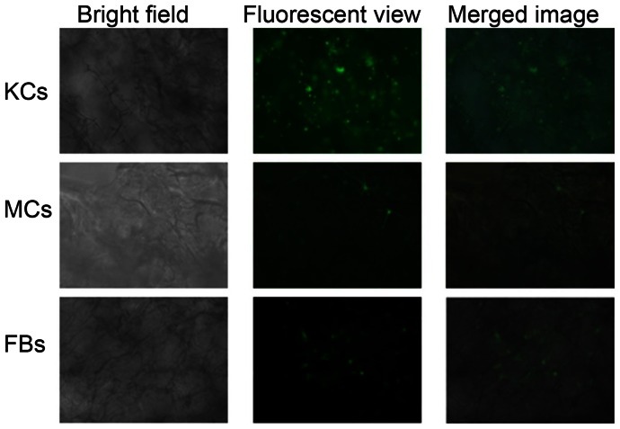
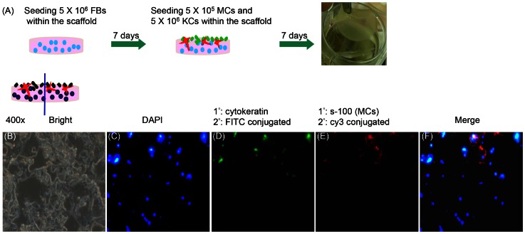
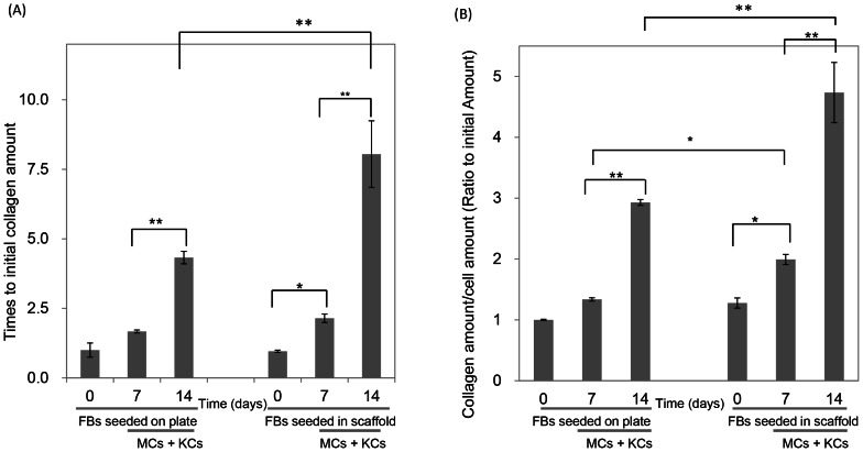
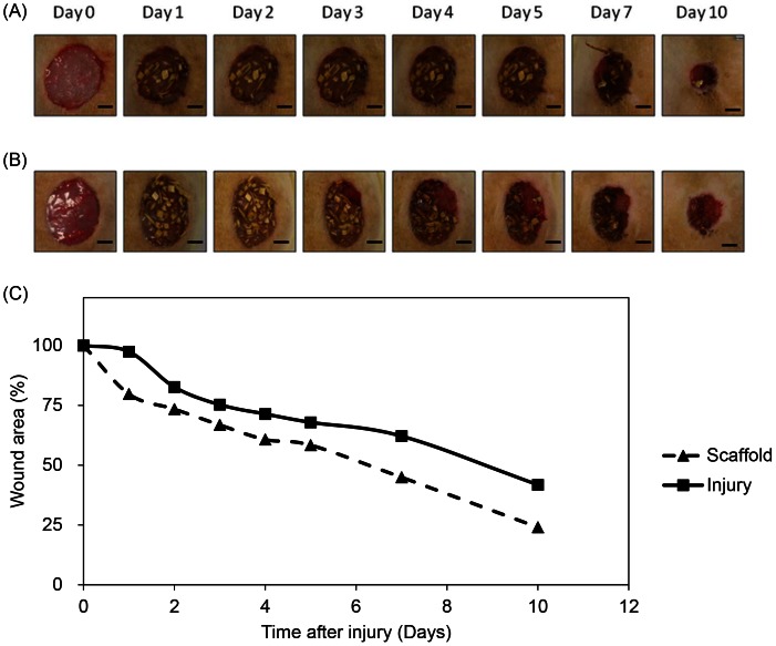
Skin wound healing is an important lifesaving issue for massive lesions. A novel porous scaffold with collagen, hyaluronic acid and gelatin was developed for skin wound repair. The swelling ratio of this developed scaffold was assayed by water absorption capacity and showed a value of over 20 g water/g dried scaffold. The scaffold was then degraded in time- and dose-dependent manners by three enzymes: lysozyme, hyaluronidase and collagenase I. The average pore diameter of the scaffold was 132.5±8.4 µm measured from SEM images. With human skin cells growing for 7 days, the SEM images showed surface fractures on the scaffold due to enzymatic digestion, indicating the biodegradable properties of this scaffold. To simulate skin distribution, the human epidermal keratinocytes, melanocytes and dermal fibroblasts were seeded on the porous scaffold and the cross-section immunofluorescent staining demonstrated normal human skin layer distributions. The collagen amount was also quantified after skin cells seeding and presented an amount 50% higher than those seeded on culture wells. The in vivo histological results showed that the scaffold ameliorated wound healing, including decreasing neutrophil infiltrates and thickening newly generated skin compared to the group without treatments.
皮肤创伤愈合是治疗大面积创伤的重要救生问题。为了进行皮肤创伤修复,开发了一种具有胶原蛋白、透明质酸和明胶的新型多孔支架。通过吸水能力测定了该开发支架的溶胀比,其值超过 20 g 水/g 干支架。然后,该支架通过三种酶:溶菌酶、透明质酸酶和胶原酶 I 以时间和剂量依赖的方式降解。从 SEM 图像测量,支架的平均孔径为 132.5±8.4 µm。用人皮肤细胞培养 7 天后,SEM 图像显示由于酶消化导致支架表面出现裂缝,表明该支架具有生物降解性。为了模拟皮肤分布,将人表皮角质形成细胞、黑素细胞和成纤维细胞接种到多孔支架上,横截面免疫荧光染色显示正常的人类皮肤层分布。在接种皮肤细胞后还定量了胶原蛋白的含量,其含量比接种在培养孔中的含量高 50%。体内组织学结果表明,与未治疗组相比,该支架可改善伤口愈合,包括减少中性粒细胞浸润和增加新生成的皮肤厚度。