Department of Chemistry and Biochemistry, South Dakota State University, Brookings, South Dakota, United States of America.
PLoS One. 2013 Jun 6;8(6):e64760. doi: 10.1371/journal.pone.0064760. Print 2013.
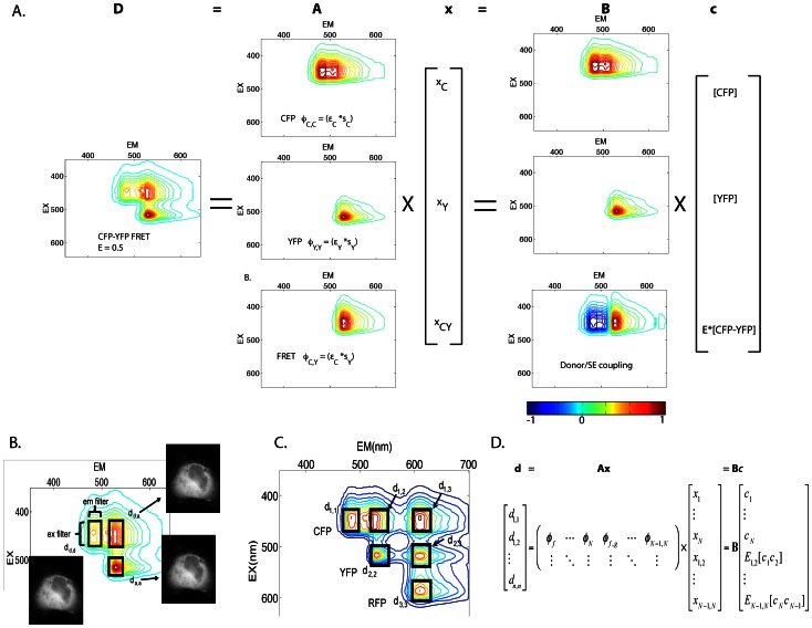
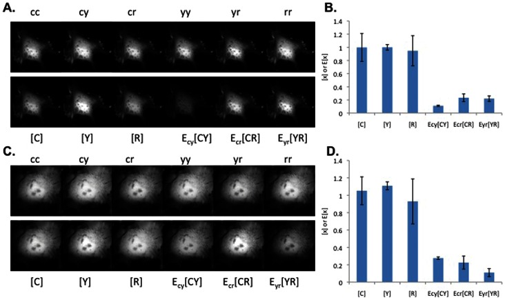
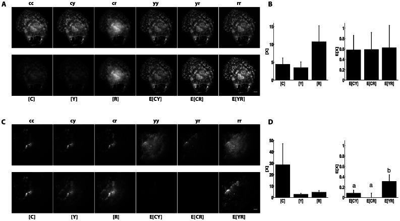
Fluorescence Resonance Energy Transfer (FRET) microscopy has emerged as a powerful tool to visualize nanoscale protein-protein interactions while capturing their microscale organization and millisecond dynamics. Recently, FRET microscopy was extended to imaging of multiple donor-acceptor pairs, thereby enabling visualization of multiple biochemical events within a single living cell. These methods require numerous equations that must be defined on a case-by-case basis. Here, we present a universal multispectral microscopy method (N-Way FRET) to enable quantitative imaging for any number of interacting and non-interacting FRET pairs. This approach redefines linear unmixing to incorporate the excitation and emission couplings created by FRET, which cannot be accounted for in conventional linear unmixing. Experiments on a three-fluorophore system using blue, yellow and red fluorescent proteins validate the method in living cells. In addition, we propose a simple linear algebra scheme for error propagation from input data to estimate the uncertainty in the computed FRET images. We demonstrate the strength of this approach by monitoring the oligomerization of three FP-tagged HIV Gag proteins whose tight association in the viral capsid is readily observed. Replacement of one FP-Gag molecule with a lipid raft-targeted FP allowed direct observation of Gag oligomerization with no association between FP-Gag and raft-targeted FP. The N-Way FRET method provides a new toolbox for capturing multiple molecular processes with high spatial and temporal resolution in living cells.
荧光共振能量转移(FRET)显微镜已成为一种强大的工具,可以可视化纳米级蛋白质-蛋白质相互作用,同时捕捉其微尺度组织和毫秒级动力学。最近,FRET 显微镜已扩展到对多个供体-受体对的成像,从而能够在单个活细胞内可视化多个生化事件。这些方法需要许多方程,这些方程必须根据具体情况进行定义。在这里,我们提出了一种通用的多光谱显微镜方法(N-Way FRET),以实现任何数量的相互作用和非相互作用的 FRET 对的定量成像。这种方法重新定义了线性解混,以纳入由 FRET 产生的激发和发射耦合,这在传统的线性解混中无法考虑。在使用蓝色、黄色和红色荧光蛋白的三荧光团系统上的实验验证了该方法在活细胞中的有效性。此外,我们提出了一种简单的线性代数方案,用于从输入数据传播误差,以估计计算出的 FRET 图像的不确定性。我们通过监测三种 FP 标记的 HIV Gag 蛋白的寡聚化来证明这种方法的优势,这些蛋白在病毒衣壳中的紧密结合很容易观察到。用靶向脂筏的 FP 取代一个 FP-Gag 分子,允许直接观察 Gag 寡聚化,而 FP-Gag 与靶向脂筏的 FP 之间没有关联。N-Way FRET 方法为在活细胞中以高时空分辨率捕获多个分子过程提供了一个新的工具箱。