Virginia Commonwealth University, Department of Biomedical Engineering, Richmond, VA, 23284, USA.
Sci Rep. 2017 Nov 15;7(1):15609. doi: 10.1038/s41598-017-15411-8.
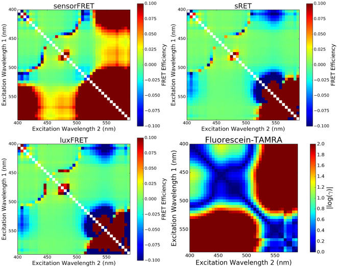
Fluorescence microscopy of FRET-based biosensors allow nanoscale interactions to be probed in living cells. This paper describes a novel approach to spectrally resolved fluorescence microscopy, termed sensorFRET, that enables quantitative measurement of FRET efficiency. This approach is an improvement on existing methods (FLIM, sRET, luxFRET, pFRET), as it does not require single fluorophore standards to be measured with every experiment and the acquisition is intensity independent, allowing the laser power to be optimized for varying levels of fluorophore expression. Additionally, it was found that all spectral based methods, including sensorFRET, fail at specific fluorophore-excitation wavelength combinations. These combinations can be determined a priori using sensorFRET, whereas other methods would give no indication of inaccuracies. This method was thoroughly validated and compared to existing methods using simulated spectra, Fluorescein and TAMRA dye mixtures as a zero FRET control, and Cerulean-Venus FRET standards as positive FRET controls. Simulations also provided a means of quantifying the uncertainty in each measurement by relating the fit residual of noisy spectra to the standard deviation of the measured FRET efficiency. As an example application, Teal-Venus force sensitive biosensors integrated into E-cadherin were used to resolve piconewton scale forces along different parts of an individual cell junction.
基于荧光共振能量转移(FRET)的生物传感器的荧光显微镜技术可用于探测活细胞中的纳米级相互作用。本文描述了一种新的荧光显微镜技术,称为 sensorFRET,它可以实现 FRET 效率的定量测量。与现有的方法(FLIM、sRET、luxFRET、pFRET)相比,这种方法是一种改进,因为它不需要对每个实验都进行单荧光团标准测量,并且采集是强度无关的,从而可以优化激光功率以适应不同水平的荧光团表达。此外,还发现所有基于光谱的方法,包括 sensorFRET,在特定的荧光团激发波长组合下都会失效。这些组合可以使用 sensorFRET 预先确定,而其他方法则不会指示出不准确的情况。该方法使用模拟光谱、Fluorescein 和 TAMRA 染料混合物作为零 FRET 对照物以及 Cerulean-Venus FRET 标准品作为正 FRET 对照物进行了全面验证和比较。模拟还通过将噪声光谱的拟合残差与测量的 FRET 效率的标准偏差相关联,为每个测量的不确定性提供了量化的方法。作为一个示例应用,整合到 E-钙黏蛋白中的 Teal-Venus 力敏感生物传感器用于解析单个细胞连接不同部位的皮牛级力。