• 文献检索
  • 文档翻译
  • 深度研究
  • 学术资讯
  • Suppr Zotero 插件Zotero 插件
  • 邀请有礼
  • 套餐&价格
  • 历史记录
应用&插件
Suppr Zotero 插件Zotero 插件浏览器插件Mac 客户端Windows 客户端微信小程序
定价
高级版会员购买积分包购买API积分包
服务
文献检索文档翻译深度研究API 文档MCP 服务
关于我们
关于 Suppr公司介绍联系我们用户协议隐私条款
关注我们

Suppr 超能文献

核心技术专利:CN118964589B侵权必究
粤ICP备2023148730 号-1Suppr @ 2026

文献检索

告别复杂PubMed语法,用中文像聊天一样搜索,搜遍4000万医学文献。AI智能推荐,让科研检索更轻松。

立即免费搜索

文件翻译

保留排版,准确专业,支持PDF/Word/PPT等文件格式,支持 12+语言互译。

免费翻译文档

深度研究

AI帮你快速写综述,25分钟生成高质量综述,智能提取关键信息,辅助科研写作。

立即免费体验

前交叉韧带和半月板损伤的诊断:膝关节屈伸的磁共振成像与关节镜检查的比较

Diagnosis of ACL and meniscal injuries: MR imaging of knee flexion versus extension compared to arthroscopy.

作者信息

Muhle Claus, Ahn Joong Mo, Dieke Constanze

机构信息

Radiology Vechta, St. Marienhospital Vechta, Marienstr. 6-8, 49377 Vechta, Germany.

出版信息

Springerplus. 2013 May 8;2(1):213. doi: 10.1186/2193-1801-2-213. Print 2013 Dec.

DOI:10.1186/2193-1801-2-213
PMID:23762813
原文链接:https://pmc.ncbi.nlm.nih.gov/articles/PMC3676741/
Abstract

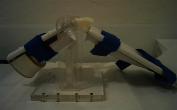
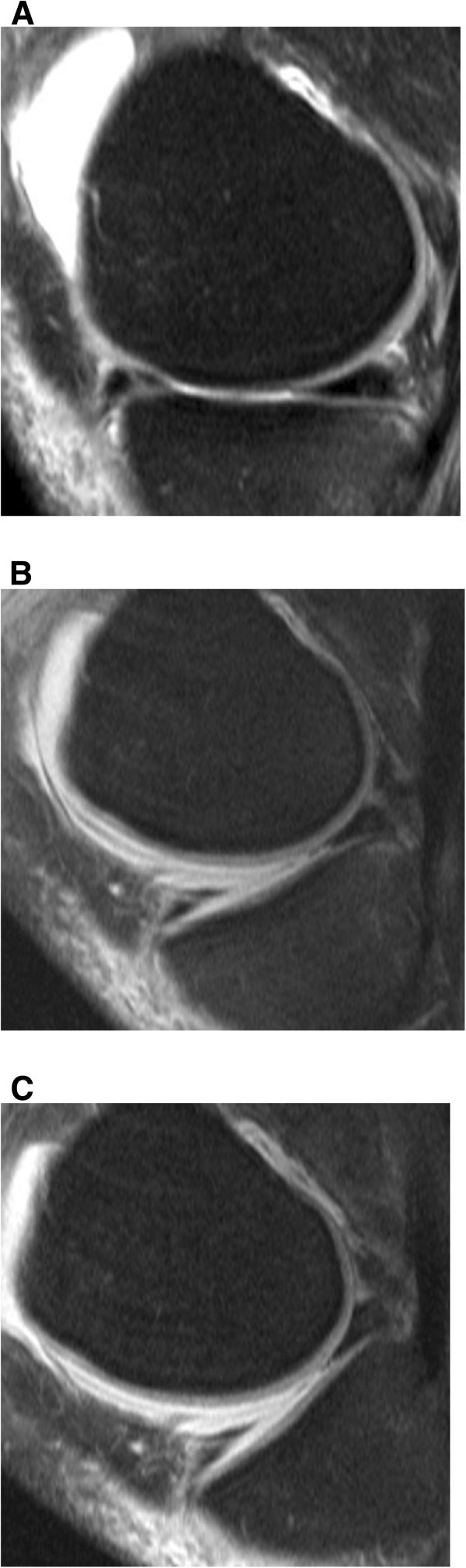
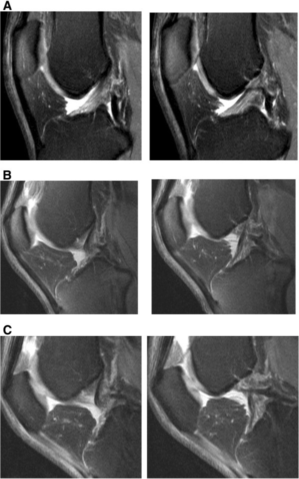
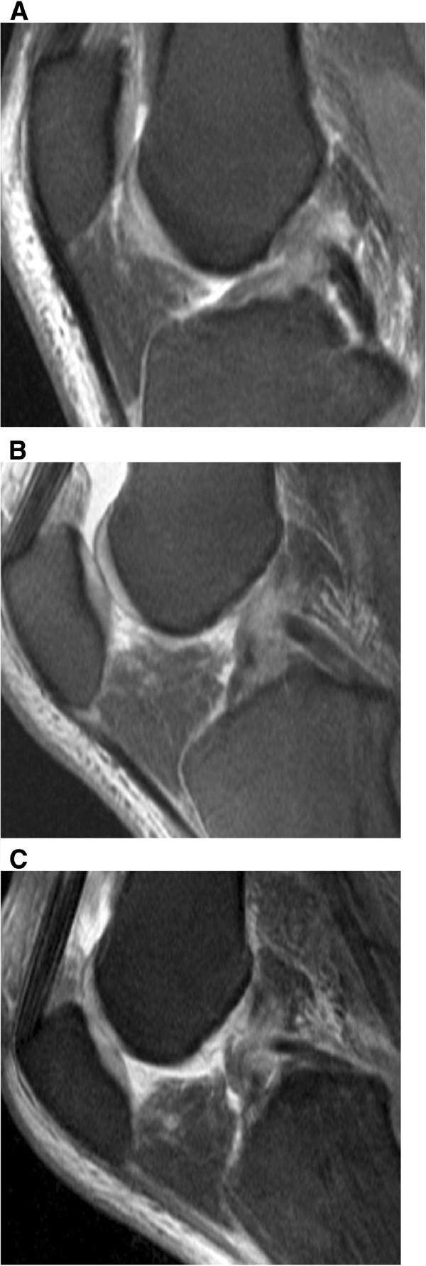
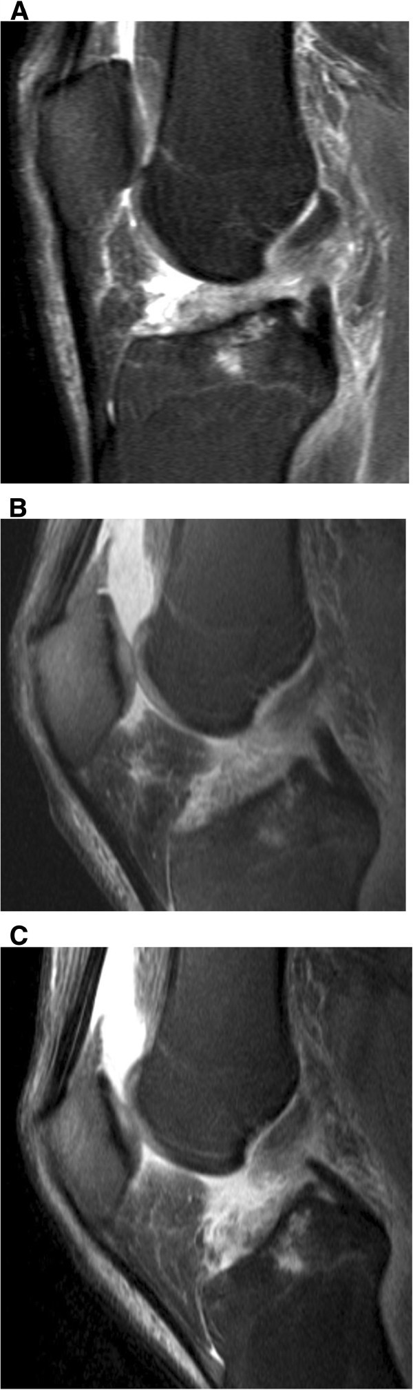
The aim of the study was to evaluate whether MR Imaging of the knee at 30° and 55° of flexion can improve the diagnosis of anterior cruciate ligament and menisci injuries compared to arthroscopy and imaging during extension of the knee joint. Knee joints from 40 patients with clinical suspicion of an anterior cruciate ligament (ACL) rupture were examined using MRI while the knee joint was either extended or flexed at 30° and 55° of knee flexion. A standard MR knee coil was used at extension, whereas at 30° and 55° of flexion a non-metallic positioning device and a flexible surface coil was placed ventral to the patella. Sagittal T2-weighted TSE sequences were acquired. In 29 of 40 patients, arthroscopy results were compared to the MRI examinations. Image quality of MRI examinations was evaluated using a three-point rating scale in a blinded fashion. Images were compared between groups and rated as better quality, same quality, or worse quality. Additionally, each angle MRI was compared to arthroscopy results. Partial ACL ruptures were diagnosed with 63% accuracy using MR imaging at 30° and 55° of knee flexion compared to 50% accuracy during knee extension. MRI imaging of complete ACL ruptures resulted in 83% accuracy of diagnosis when imaged at 30° flexion, 93% accuracy at 55° flexion, and 83% accuracy at extension. The accuracy of diagnosing medial meniscus lesions was 73% at extension, 64% at 30° flexion and 73% at 55° of flexion. MR imaging was only able to diagnose lateral meniscus tears with 55% accuracy in all three knee positions. The diagnosis of meniscal tears was more difficult due to small peripheral tears. The improved results in the diagnosis of ACL tears in response to 30° flexion and in particular in response to 55° flexion were based on the fact that the anterior cruciate ligament moved further away from the intercondylar roof with increased knee flexion. During flexion the ligament tension decreased, which causes the anterior cruciate ligament to have cylindrical shape and therefore made visualization of the injury easier. In conclusion, MR Imaging of the knee at 55° of flexion and less at 30° of flexion allows an improved diagnosis of injuries to the anterior cruciate ligament as compared to MRI examinations at extension. The diagnosis of meniscal injuries, however, was not superior at both flexion positions compared to commonly performed examinations at knee extension.

摘要

本研究的目的是评估膝关节在30°和55°屈曲位时的磁共振成像(MRI)与关节镜检查及膝关节伸展位成像相比,是否能改善前交叉韧带和半月板损伤的诊断。对40例临床怀疑前交叉韧带(ACL)断裂的患者的膝关节进行MRI检查,检查时膝关节处于伸展位或分别屈曲30°和55°。膝关节伸展位时使用标准的膝关节MRI线圈,而在屈曲30°和55°时,在髌骨前方放置一个非金属定位装置和一个柔性表面线圈。采集矢状面T2加权快速自旋回波序列。在40例患者中的29例中,将关节镜检查结果与MRI检查结果进行了比较。采用三点评分量表以盲法评估MRI检查的图像质量。对不同组的图像进行比较,并评为质量更好、质量相同或质量更差。此外,将每个角度的MRI与关节镜检查结果进行比较。与膝关节伸展位时50%的准确率相比,膝关节在30°和55°屈曲位时采用MRI诊断部分ACL断裂的准确率为63%。完全ACL断裂的MRI成像在膝关节屈曲30°时诊断准确率为83%,屈曲55°时为93%,伸展位时为83%。诊断内侧半月板损伤在伸展位时的准确率为73%,屈曲30°时为64%,屈曲55°时为73%。在所有三个膝关节位置,MR成像诊断外侧半月板撕裂的准确率仅为55%。由于半月板周边小撕裂,半月板撕裂的诊断较为困难。在诊断ACL撕裂方面,30°屈曲尤其是55°屈曲时结果有所改善,这是基于随着膝关节屈曲增加,前交叉韧带与髁间顶的距离进一步增大这一事实。在屈曲过程中韧带张力降低,这使得前交叉韧带呈圆柱形,因此更容易观察到损伤情况。总之,与膝关节伸展位的MRI检查相比,膝关节在55°屈曲位及较少程度的30°屈曲位时的MRI检查能改善前交叉韧带损伤的诊断。然而,与膝关节伸展位的常规检查相比,在这两个屈曲位时半月板损伤的诊断并无优势。

https://cdn.ncbi.nlm.nih.gov/pmc/blobs/6041/3676741/529e6857f530/40064_2012_309_Fig5_HTML.jpg
https://cdn.ncbi.nlm.nih.gov/pmc/blobs/6041/3676741/16252e83b1ad/40064_2012_309_Fig1_HTML.jpg
https://cdn.ncbi.nlm.nih.gov/pmc/blobs/6041/3676741/5e96cb19f7df/40064_2012_309_Fig2_HTML.jpg
https://cdn.ncbi.nlm.nih.gov/pmc/blobs/6041/3676741/3c5f56a141d0/40064_2012_309_Fig3_HTML.jpg
https://cdn.ncbi.nlm.nih.gov/pmc/blobs/6041/3676741/7eb2dfceee5a/40064_2012_309_Fig4_HTML.jpg
https://cdn.ncbi.nlm.nih.gov/pmc/blobs/6041/3676741/529e6857f530/40064_2012_309_Fig5_HTML.jpg

相似文献

1
Diagnosis of ACL and meniscal injuries: MR imaging of knee flexion versus extension compared to arthroscopy.前交叉韧带和半月板损伤的诊断:膝关节屈伸的磁共振成像与关节镜检查的比较
Springerplus. 2013 May 8;2(1):213. doi: 10.1186/2193-1801-2-213. Print 2013 Dec.
2
Meniscal tears missed on MR imaging: relationship to meniscal tear patterns and anterior cruciate ligament tears.磁共振成像漏诊的半月板撕裂:与半月板撕裂模式及前交叉韧带撕裂的关系
AJR Am J Roentgenol. 1994 Apr;162(4):905-11. doi: 10.2214/ajr.162.4.8141016.
3
The value of the sagittal-oblique MRI technique for injuries of the anterior cruciate ligament in the knee.MRI 矢状斜位技术在膝关节前交叉韧带损伤中的价值。
Radiol Oncol. 2013 Mar;47(1):19-25. doi: 10.2478/raon-2013-0006. Epub 2013 Feb 1.
4
Partial meniscectomy does not affect the biomechanics of anterior cruciate ligament reconstructed knee with a lateral posterior meniscal root tear.部分半月板切除术不会影响外侧后半月板根部撕裂的前交叉韧带重建膝关节的生物力学。
Knee Surg Sports Traumatol Arthrosc. 2020 Nov;28(11):3481-3487. doi: 10.1007/s00167-020-06209-9. Epub 2020 Sep 5.
5
Meniscal repair concurrent with anterior cruciate ligament reconstruction restores posterior shift of the medial meniscus in the knee-flexed position.半月板修复术同期进行前交叉韧带重建术可恢复膝关节屈膝位时内侧半月板的后移。
Knee Surg Sports Traumatol Arthrosc. 2019 Feb;27(2):361-368. doi: 10.1007/s00167-018-5157-2. Epub 2018 Sep 24.
6
Reliability of magnetic resonance imaging in evaluating meniscal and cartilage injuries in anterior cruciate ligament-deficient knees.磁共振成像在评估前交叉韧带损伤膝关节半月板和软骨损伤中的可靠性。
Knee Surg Sports Traumatol Arthrosc. 2017 Feb;25(2):411-417. doi: 10.1007/s00167-016-4211-1. Epub 2016 Jun 24.
7
Prospective comparison of 1.5 and 3.0-T MRI for evaluating the knee menisci and ACL.1.5T 和 3.0T MRI 评估膝关节半月板和 ACL 的前瞻性比较。
J Bone Joint Surg Am. 2013 May 15;95(10):916-24. doi: 10.2106/JBJS.L.01195.
8
High-resolution oblique coronal MRI at optimal flexed-knee angle: a novel imaging method for enhanced anterior cruciate ligament tear diagnosis.高分辨率斜冠状 MRI 在最佳屈膝角度:增强前交叉韧带撕裂诊断的新成像方法。
J Orthop Surg Res. 2024 Aug 1;19(1):456. doi: 10.1186/s13018-024-04956-w.
9
Slightly flexed knee position within a standard knee coil: MR delineation of the anterior cruciate ligament.在标准膝关节线圈内保持膝关节轻度屈曲位:前交叉韧带的磁共振成像描绘
Eur Radiol. 1998;8(1):113-5. doi: 10.1007/s003300050349.
10
Knee extension and flexion: MR delineation of normal and torn anterior cruciate ligaments.
J Comput Assist Tomogr. 1996 Mar-Apr;20(2):322-7. doi: 10.1097/00004728-199603000-00030.

引用本文的文献

1
High-resolution oblique coronal MRI at optimal flexed-knee angle: a novel imaging method for enhanced anterior cruciate ligament tear diagnosis.高分辨率斜冠状 MRI 在最佳屈膝角度:增强前交叉韧带撕裂诊断的新成像方法。
J Orthop Surg Res. 2024 Aug 1;19(1):456. doi: 10.1186/s13018-024-04956-w.
2
Risk factors for false positive and false negative MRI in diagnosing medial and lateral meniscal tears with concomitant ACL injury.在诊断伴有前交叉韧带损伤的内侧和外侧半月板撕裂时,MRI假阳性和假阴性的危险因素。
Skeletal Radiol. 2025 Feb;54(2):303-315. doi: 10.1007/s00256-024-04745-w. Epub 2024 Jul 8.
3
How Many Bundles Does the Anterior Cruciate Ligament Consist of? A Case Report.

本文引用的文献

1
Factors used to determine return to unrestricted sports activities after anterior cruciate ligament reconstruction.前交叉韧带重建术后恢复非限制型运动活动的决定因素。
Arthroscopy. 2011 Dec;27(12):1697-705. doi: 10.1016/j.arthro.2011.09.009.
2
[MRI based volumetric assessment of knee cartilage after ACL-reconstruction, correlated with qualitative morphologic changes in the joint and with clinical outcome. Is there evidence for early posttraumatic degeneration?].[基于MRI的前交叉韧带重建术后膝关节软骨容积评估,与关节定性形态学变化及临床结果相关。是否有创伤后早期退变的证据?]
Rofo. 2011 Dec;183(12):1138-44. doi: 10.1055/s-0031-1281685. Epub 2011 Sep 28.
3
前交叉韧带由多少束组成?一例病例报告。
Sisli Etfal Hastan Tip Bul. 2023 Dec 29;57(4):557-562. doi: 10.14744/SEMB.2022.12844. eCollection 2023.
4
Meniscal extrusion: risk factors and diagnostic tools to predict early osteoarthritis.半月板挤出:预测早期骨关节炎的危险因素和诊断工具。
Orthop Rev (Pavia). 2023 May 13;15:74881. doi: 10.52965/001c.74881. eCollection 2023.
5
Standard MRI May Not Predict Specific Acute Anterior Cruciate Ligament Rupture Characteristics.标准磁共振成像可能无法预测特定的急性前交叉韧带断裂特征。
Orthop J Sports Med. 2021 Mar 29;9(3):2325967121992472. doi: 10.1177/2325967121992472. eCollection 2021 Mar.
6
Non-clinical assessment design of autologous chondrocyte implantation products.自体软骨细胞植入产品的非临床评估设计
Regen Ther. 2015 Jul 13;1:98-108. doi: 10.1016/j.reth.2015.06.003. eCollection 2015 Jun.
7
Unlocking a locked knee by ultrasound guided anesthetic injection.
Radiol Case Rep. 2019 Feb 25;14(5):548-550. doi: 10.1016/j.radcr.2019.01.013. eCollection 2019 May.
8
Use of MRI by radiologists and orthopaedic surgeons to detect intra-articular injuries of the knee.放射科医生和骨科医生使用磁共振成像(MRI)检测膝关节的关节内损伤。
Rev Bras Ortop. 2017 Jan 17;53(1):28-32. doi: 10.1016/j.rboe.2016.12.013. eCollection 2018 Jan-Feb.
9
Does anatomic single-bundle ACL reconstruction using hamstring autograft produce anterolateral meniscal root tearing?使用自体腘绳肌腱进行解剖单束前交叉韧带重建会导致前外侧半月板根部撕裂吗?
J Exp Orthop. 2017 Dec;4(1):17. doi: 10.1186/s40634-017-0093-5. Epub 2017 May 22.
10
A dynamic study of the anterior cruciate ligament of the knee using an open MRI.使用开放式磁共振成像对膝关节前交叉韧带进行的动态研究。
Surg Radiol Anat. 2017 Mar;39(3):307-314. doi: 10.1007/s00276-016-1730-x. Epub 2016 Aug 11.
MRI of the knee ligaments and menisci: comparison of isotropic-resolution 3D and conventional 2D fast spin-echo sequences at 3 T.
膝关节韧带和半月板的 MRI:3T 下各向同性分辨率 3D 与传统 2D 快速自旋回波序列的比较。
AJR Am J Roentgenol. 2011 Aug;197(2):442-50. doi: 10.2214/AJR.10.5709.
4
Stable or unstable tear of the anterior cruciate ligament of the knee: an MR diagnosis?膝关节前交叉韧带稳定或不稳定撕裂:MR 诊断?
Skeletal Radiol. 2012 Mar;41(3):273-80. doi: 10.1007/s00256-011-1169-4. Epub 2011 Apr 19.
5
[Bucket handle tears].
Rofo. 2011 Feb;183(2):91-4. doi: 10.1055/s-0030-1270803. Epub 2011 Feb 8.
6
Three tesla magnetic resonance imaging of the anterior cruciate ligament of the knee: can we differentiate complete from partial tears?膝关节前交叉韧带 3T 磁共振成像:能否区分完全撕裂与部分撕裂?
Skeletal Radiol. 2011 Jun;40(6):701-7. doi: 10.1007/s00256-010-1044-8. Epub 2010 Oct 8.
7
Magnetic resonance imaging of cruciate ligament injuries of the knee.膝关节十字韧带损伤的磁共振成像。
Can Assoc Radiol J. 2010 Apr;61(2):80-9. doi: 10.1016/j.carj.2009.11.003. Epub 2010 Jan 29.
8
Tibiofemoral kinematics following successful anterior cruciate ligament reconstruction using dynamic multiple resonance imaging.使用动态多重共振成像技术对成功进行前交叉韧带重建后的胫股关节运动学研究
Am J Sports Med. 2004 Jun;32(4):984-92. doi: 10.1177/0363546503261702.
9
MR imaging of meniscal and cruciate ligament injuries.
Magn Reson Imaging Clin N Am. 2003 May;11(2):283-93. doi: 10.1016/s1064-9689(03)00028-x.
10
MR imaging of the flexed knee: comparison to the extended knee in delineation of meniscal lesions.
Eur Radiol. 2000;10(11):1824-7. doi: 10.1007/s003300000442.