Azar Jimmy C, Busch Christer, Carlbom Ingrid B
Department of Information Technology, Centre for Image Analysis, Uppsala University, Uppsala, Sweden.
J Pathol Inform. 2013 Mar 30;4(Suppl):S11. doi: 10.4103/2153-3539.109869. Print 2013.
A methodology for quantitative comparison of histological stains based on their classification and clustering performance, which may facilitate the choice of histological stains for automatic pattern and image analysis.
Machine learning and image analysis are becoming increasingly important in pathology applications for automatic analysis of histological tissue samples. Pathologists rely on multiple, contrasting stains to analyze tissue samples, but histological stains are developed for visual analysis and are not always ideal for automatic analysis.
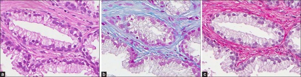
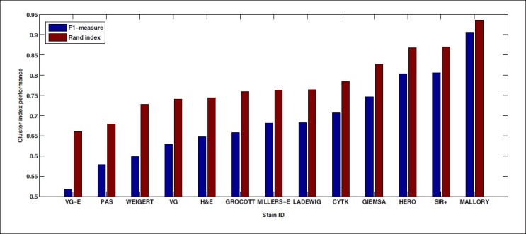
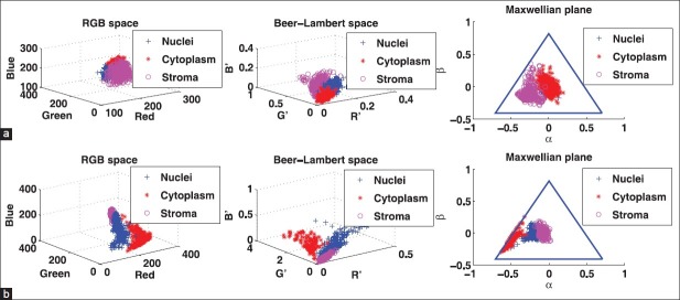
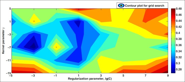
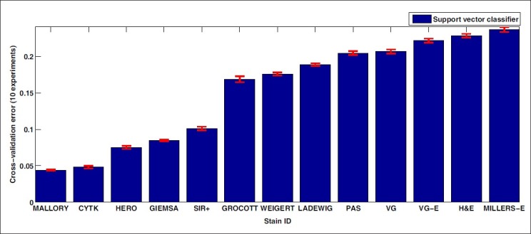
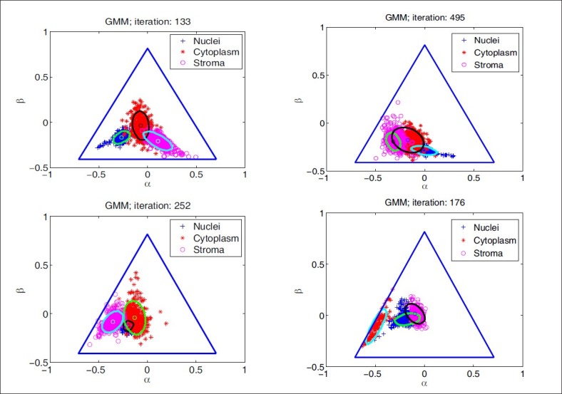
Thirteen different histological stains were used to stain adjacent prostate tissue sections from radical prostatectomies. We evaluate the stains for both supervised and unsupervised classification of stain/tissue combinations. For supervised classification we measure the error rate of nonlinear support vector machines, and for unsupervised classification we use the Rand index and the F-measure to assess the clustering results of a Gaussian mixture model based on expectation-maximization. Finally, we investigate class separability measures based on scatter criteria.
A methodology for quantitative evaluation of histological stains in terms of their classification and clustering efficacy that aims at improving segmentation and color decomposition. We demonstrate that for a specific tissue type, certain stains perform consistently better than others according to objective error criteria.
The choice of histological stain for automatic analysis must be based on its classification and clustering performance, which are indicators of the performance of automatic segmentation of tissue into morphological components, which in turn may be the basis for diagnosis.
一种基于组织学染色的分类和聚类性能进行定量比较的方法,这可能有助于为自动模式和图像分析选择组织学染色。
机器学习和图像分析在病理学应用中对组织学组织样本的自动分析变得越来越重要。病理学家依靠多种对比染色来分析组织样本,但组织学染色是为视觉分析而开发的,并不总是适合自动分析。
使用13种不同的组织学染色对前列腺癌根治术的相邻前列腺组织切片进行染色。我们评估这些染色在染色/组织组合的监督和非监督分类方面的性能。对于监督分类,我们测量非线性支持向量机的错误率,对于非监督分类,我们使用兰德指数和F值来评估基于期望最大化的高斯混合模型的聚类结果。最后,我们研究基于散射标准的类可分性度量。
一种基于分类和聚类效果对组织学染色进行定量评估的方法,旨在改善分割和颜色分解。我们证明,对于特定的组织类型,根据客观错误标准,某些染色的表现始终优于其他染色。
用于自动分析的组织学染色的选择必须基于其分类和聚类性能,这些性能是将组织自动分割为形态学成分的性能指标,而这反过来可能是诊断的基础。