• 文献检索
  • 文档翻译
  • 深度研究
  • 学术资讯
  • Suppr Zotero 插件Zotero 插件
  • 邀请有礼
  • 套餐&价格
  • 历史记录
应用&插件
Suppr Zotero 插件Zotero 插件浏览器插件Mac 客户端Windows 客户端微信小程序
定价
高级版会员购买积分包购买API积分包
服务
文献检索文档翻译深度研究API 文档MCP 服务
关于我们
关于 Suppr公司介绍联系我们用户协议隐私条款
关注我们

Suppr 超能文献

核心技术专利:CN118964589B侵权必究
粤ICP备2023148730 号-1Suppr @ 2026

文献检索

告别复杂PubMed语法,用中文像聊天一样搜索,搜遍4000万医学文献。AI智能推荐,让科研检索更轻松。

立即免费搜索

文件翻译

保留排版,准确专业,支持PDF/Word/PPT等文件格式,支持 12+语言互译。

免费翻译文档

深度研究

AI帮你快速写综述,25分钟生成高质量综述,智能提取关键信息,辅助科研写作。

立即免费体验

基于模式分类的动脉粥样硬化组织学自动分割

Automated segmentation of atherosclerotic histology based on pattern classification.

作者信息

van Engelen Arna, Niessen Wiro J, Klein Stefan, Groen Harald C, van Gaalen Kim, Verhagen Hence J, Wentzel Jolanda J, van der Lugt Aad, de Bruijne Marleen

机构信息

Biomedical Imaging Group Rotterdam, Department of Medical Informatics and Radiology, Erasmus Medical Centre, Rotterdam, Netherlands.

出版信息

J Pathol Inform. 2013 Mar 30;4(Suppl):S3. doi: 10.4103/2153-3539.109844. Print 2013.

DOI:10.4103/2153-3539.109844
PMID:23766939
原文链接:https://pmc.ncbi.nlm.nih.gov/articles/PMC3678743/
Abstract

BACKGROUND

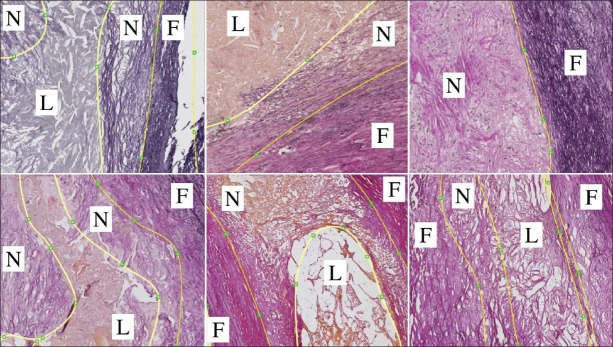
Histology sections provide accurate information on atherosclerotic plaque composition, and are used in various applications. To our knowledge, no automated systems for plaque component segmentation in histology sections currently exist.

MATERIALS AND METHODS

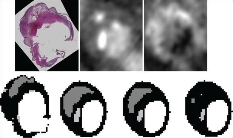
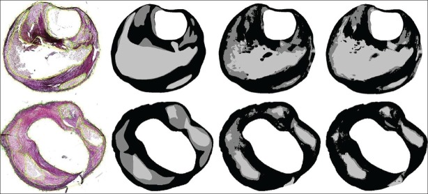
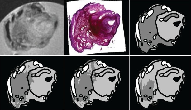
We perform pixel-wise classification of fibrous, lipid, and necrotic tissue in Elastica Von Gieson-stained histology sections, using features based on color channel intensity and local image texture and structure. We compare an approach where we train on independent data to an approach where we train on one or two sections per specimen in order to segment the remaining sections. We evaluate the results on segmentation accuracy in histology, and we use the obtained histology segmentations to train plaque component classification methods in ex vivo Magnetic resonance imaging (MRI) and in vivo MRI and computed tomography (CT).

RESULTS

In leave-one-specimen-out experiments on 176 histology slices of 13 plaques, a pixel-wise accuracy of 75.7 ± 6.8% was obtained. This increased to 77.6 ± 6.5% when two manually annotated slices of the specimen to be segmented were used for training. Rank correlations of relative component volumes with manually annotated volumes were high in this situation (P = 0.82-0.98). Using the obtained histology segmentations to train plaque component classification methods in ex vivo MRI and in vivo MRI and CT resulted in similar image segmentations for training on the automated histology segmentations as for training on a fully manual ground truth. The size of the lipid-rich necrotic core was significantly smaller when training on fully automated histology segmentations than when manually annotated histology sections were used. This difference was reduced and not statistically significant when one or two slices per section were manually annotated for histology segmentation.

CONCLUSIONS

Good histology segmentations can be obtained by automated segmentation, which show good correlations with ground truth volumes. In addition, these can be used to develop segmentation methods in other imaging modalities. Accuracy increases when one or two sections of the same specimen are used for training, which requires a limited amount of user interaction in practice.

摘要

背景

组织学切片可提供有关动脉粥样硬化斑块成分的准确信息,并应用于各种领域。据我们所知,目前尚无用于组织学切片中斑块成分分割的自动化系统。

材料与方法

我们利用基于颜色通道强度以及局部图像纹理和结构的特征,对弹性冯吉森染色组织学切片中的纤维、脂质和坏死组织进行逐像素分类。我们将在独立数据上进行训练的方法与在每个标本的一两个切片上进行训练以分割其余切片的方法进行比较。我们评估组织学分割准确性的结果,并使用获得的组织学分割结果来训练离体磁共振成像(MRI)、活体MRI和计算机断层扫描(CT)中的斑块成分分类方法。

结果

在对13个斑块的176个组织学切片进行留一标本法实验中,获得了75.7±6.8%的逐像素准确率。当使用待分割标本的两个手动标注切片进行训练时,该准确率提高到了77.6±6.5%。在这种情况下,相对成分体积与手动标注体积的等级相关性很高(P = 0.82 - 0.98)。使用获得的组织学分割结果来训练离体MRI、活体MRI和CT中的斑块成分分类方法,对于基于自动化组织学分割进行训练所得到的图像分割,与基于完全手动的真实情况进行训练所得到的相似。与使用手动标注的组织学切片进行训练相比,基于完全自动化组织学分割进行训练时,富含脂质的坏死核心的大小显著更小。当每个标本手动标注一两个切片用于组织学分割时,这种差异减小且无统计学意义。

结论

通过自动分割可获得良好的组织学分割结果,其与真实体积显示出良好的相关性。此外,这些结果可用于开发其他成像模态下的分割方法。当使用同一标本的一两个切片进行训练时,准确率会提高,而这在实际操作中只需有限的用户交互。

https://cdn.ncbi.nlm.nih.gov/pmc/blobs/a30b/3678743/e8216fad8787/JPI-4-3-g008.jpg
https://cdn.ncbi.nlm.nih.gov/pmc/blobs/a30b/3678743/5fa05d8aa895/JPI-4-3-g001.jpg
https://cdn.ncbi.nlm.nih.gov/pmc/blobs/a30b/3678743/2d304b5c20a1/JPI-4-3-g004.jpg
https://cdn.ncbi.nlm.nih.gov/pmc/blobs/a30b/3678743/a87c4efa3c71/JPI-4-3-g006.jpg
https://cdn.ncbi.nlm.nih.gov/pmc/blobs/a30b/3678743/e8216fad8787/JPI-4-3-g008.jpg

相似文献

1
Automated segmentation of atherosclerotic histology based on pattern classification.基于模式分类的动脉粥样硬化组织学自动分割
J Pathol Inform. 2013 Mar 30;4(Suppl):S3. doi: 10.4103/2153-3539.109844. Print 2013.
2
Carotid atherosclerotic plaque segmentation in multi-weighted MRI using a two-stage neural network: advantages of training with high-resolution imaging and histology.使用两阶段神经网络在多加权磁共振成像中进行颈动脉粥样硬化斑块分割:高分辨率成像和组织学训练的优势
Front Cardiovasc Med. 2023 May 24;10:1127653. doi: 10.3389/fcvm.2023.1127653. eCollection 2023.
3
Atherosclerotic plaque component segmentation in combined carotid MRI and CTA data incorporating class label uncertainty.结合颈动脉MRI和CTA数据并纳入类别标签不确定性的动脉粥样硬化斑块成分分割
PLoS One. 2014 Apr 24;9(4):e94840. doi: 10.1371/journal.pone.0094840. eCollection 2014.
4
Deep learning of the sectional appearances of 3D CT images for anatomical structure segmentation based on an FCN voting method.基于 FCN 投票方法的三维 CT 图像节段外观的深度学习用于解剖结构分割。
Med Phys. 2017 Oct;44(10):5221-5233. doi: 10.1002/mp.12480. Epub 2017 Aug 31.
5
Segmentation evaluation with sparse ground truth data: Simulating true segmentations as perfect/imperfect as those generated by humans.使用稀疏真实数据进行分割评估:将真实分割模拟为与人类生成的分割一样完美/不完美。
Med Image Anal. 2021 Apr;69:101980. doi: 10.1016/j.media.2021.101980. Epub 2021 Jan 26.
6
Multi-feature-based plaque characterization in ex vivo MRI trained by registration to 3D histology.基于多特征的斑块特征描述,通过与 3D 组织学配准的体外 MRI 训练得到。
Phys Med Biol. 2012 Jan 7;57(1):241-56. doi: 10.1088/0031-9155/57/1/241.
7
Multi-atlas segmentation of the whole hippocampus and subfields using multiple automatically generated templates.使用多个自动生成的模板对整个海马体及其子区进行多图谱分割。
Neuroimage. 2014 Nov 1;101:494-512. doi: 10.1016/j.neuroimage.2014.04.054. Epub 2014 Apr 29.
8
Channel width optimized neural networks for liver and vessel segmentation in liver iron quantification.用于肝脏铁定量中肝脏和血管分割的通道宽度优化神经网络。
Comput Biol Med. 2020 Jul;122:103798. doi: 10.1016/j.compbiomed.2020.103798. Epub 2020 May 16.
9
An investigation of the effect of fat suppression and dimensionality on the accuracy of breast MRI segmentation using U-nets.利用 U-Nets 研究脂肪抑制和维度对乳腺 MRI 分割准确性的影响。
Med Phys. 2019 Mar;46(3):1230-1244. doi: 10.1002/mp.13375. Epub 2019 Feb 4.
10
Concurrent segmentation of the prostate on MRI and CT via linked statistical shape models for radiotherapy planning.基于链接统计形状模型的 MRI 和 CT 前列腺同步分割用于放射治疗计划。
Med Phys. 2012 Apr;39(4):2214-28. doi: 10.1118/1.3696376.

引用本文的文献

1
An alternative reference space for H&E color normalization.苏木精-伊红染色颜色标准化的另一种参考空间。
PLoS One. 2017 Mar 29;12(3):e0174489. doi: 10.1371/journal.pone.0174489. eCollection 2017.

本文引用的文献

1
Multi-feature-based plaque characterization in ex vivo MRI trained by registration to 3D histology.基于多特征的斑块特征描述,通过与 3D 组织学配准的体外 MRI 训练得到。
Phys Med Biol. 2012 Jan 7;57(1):241-56. doi: 10.1088/0031-9155/57/1/241.
2
Effects of intima stiffness and plaque morphology on peak cap stress.内膜硬度和斑块形态对峰值应力的影响。
Biomed Eng Online. 2011 Apr 8;10:25. doi: 10.1186/1475-925X-10-25.
3
Histopathological image analysis: a review.组织病理学图像分析:综述。
IEEE Rev Biomed Eng. 2009;2:147-71. doi: 10.1109/RBME.2009.2034865. Epub 2009 Oct 30.
4
Three-dimensional registration of histology of human atherosclerotic carotid plaques to in-vivo imaging.人体动脉粥样硬化颈动脉斑块的组织学与体内成像的三维配准。
J Biomech. 2010 Aug 10;43(11):2087-92. doi: 10.1016/j.jbiomech.2010.04.005. Epub 2010 May 4.
5
Automated in vivo segmentation of carotid plaque MRI with Morphology-Enhanced probability maps.利用形态学增强概率图对颈动脉斑块磁共振成像进行自动体内分割。
Magn Reson Med. 2006 Mar;55(3):659-68. doi: 10.1002/mrm.20814.
6
Reproducibility of histological assessment of carotid plaque: implications for studies of carotid imaging.颈动脉斑块组织学评估的可重复性:对颈动脉成像研究的影响
Cerebrovasc Dis. 2004;18(2):117-23. doi: 10.1159/000079259. Epub 2004 Jun 22.
7
Quantitative assessment of carotid plaque composition using multicontrast MRI and registered histology.使用多对比磁共振成像和配准组织学对颈动脉斑块成分进行定量评估。
Magn Reson Med. 2003 Dec;50(6):1199-208. doi: 10.1002/mrm.10618.
8
The relationship between carotid plaque composition, plaque morphology, and neurologic symptoms.颈动脉斑块成分、斑块形态与神经症状之间的关系。
J Surg Res. 1995 Mar;58(3):330-6. doi: 10.1006/jsre.1995.1051.