Zarella Mark D, Yeoh Chan, Breen David E, Garcia Fernando U
Department of Pathology & Laboratory Medicine, Drexel University, Philadelphia, PA, United States of America.
Department of Electrical & Computer Engineering, Drexel University, Philadelphia, PA, United States of America.
PLoS One. 2017 Mar 29;12(3):e0174489. doi: 10.1371/journal.pone.0174489. eCollection 2017.
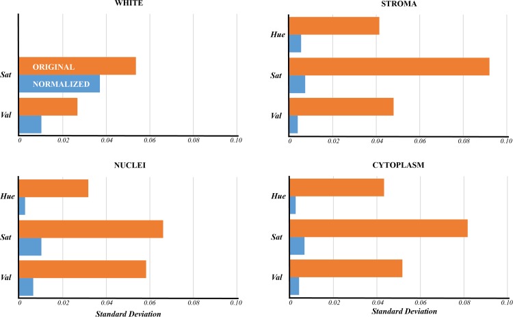
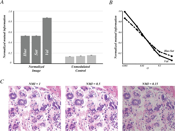
Digital imaging of H&E stained slides has enabled the application of image processing to support pathology workflows. Potential applications include computer-aided diagnostics, advanced quantification tools, and innovative visualization platforms. However, the intrinsic variability of biological tissue and the vast differences in tissue preparation protocols often lead to significant image variability that can hamper the effectiveness of these computational tools. We developed an alternative representation for H&E images that operates within a space that is more amenable to many of these image processing tools. The algorithm to derive this representation operates by exploiting the correlation between color and the spatial properties of the biological structures present in most H&E images. In this way, images are transformed into a structure-centric space in which images are segregated into tissue structure channels. We demonstrate that this framework can be extended to achieve color normalization, effectively reducing inter-slide variability.
苏木精-伊红(H&E)染色玻片的数字成像使得图像处理能够应用于支持病理学工作流程。潜在的应用包括计算机辅助诊断、先进的定量工具和创新的可视化平台。然而,生物组织的内在变异性以及组织制备方案的巨大差异常常导致显著的图像变异性,这可能会妨碍这些计算工具的有效性。我们开发了一种用于H&E图像的替代表示方法,该方法在一个更适合许多此类图像处理工具的空间内运行。导出这种表示的算法通过利用大多数H&E图像中颜色与生物结构空间属性之间的相关性来运行。通过这种方式,图像被转换到一个以结构为中心的空间,在该空间中图像被分离成组织结构通道。我们证明这个框架可以扩展以实现颜色归一化,有效减少玻片间的变异性。