• 文献检索
  • 文档翻译
  • 深度研究
  • 学术资讯
  • Suppr Zotero 插件Zotero 插件
  • 邀请有礼
  • 套餐&价格
  • 历史记录
应用&插件
Suppr Zotero 插件Zotero 插件浏览器插件Mac 客户端Windows 客户端微信小程序
定价
高级版会员购买积分包购买API积分包
服务
文献检索文档翻译深度研究API 文档MCP 服务
关于我们
关于 Suppr公司介绍联系我们用户协议隐私条款
关注我们

Suppr 超能文献

核心技术专利:CN118964589B侵权必究
粤ICP备2023148730 号-1Suppr @ 2026

文献检索

告别复杂PubMed语法,用中文像聊天一样搜索,搜遍4000万医学文献。AI智能推荐,让科研检索更轻松。

立即免费搜索

文件翻译

保留排版,准确专业,支持PDF/Word/PPT等文件格式,支持 12+语言互译。

免费翻译文档

深度研究

AI帮你快速写综述,25分钟生成高质量综述,智能提取关键信息,辅助科研写作。

立即免费体验

通过形态学尺度量化局部异质性:区分肿瘤区域和基质区域。

Quantifying local heterogeneity via morphologic scale: Distinguishing tumoral from stromal regions.

作者信息

Janowczyk Andrew, Chandran Sharat, Madabhushi Anant

机构信息

Department of Computer Science, IIT Bombay, India, USA ; Department of Biomedical Engineering, Case Western Reserve University, USA.

出版信息

J Pathol Inform. 2013 Mar 30;4(Suppl):S8. doi: 10.4103/2153-3539.109865. Print 2013.

DOI:10.4103/2153-3539.109865
PMID:23766944
原文链接:https://pmc.ncbi.nlm.nih.gov/articles/PMC3678744/
Abstract

INTRODUCTION

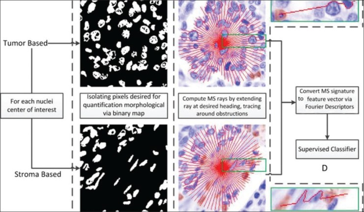
The notion of local scale was introduced to characterize varying levels of image detail so that localized image processing tasks could be performed while simultaneously yielding a globally optimal result. In this paper, we have presented the methodological framework for a novel locally adaptive scale definition, morphologic scale (MS), which is different from extant local scale definitions in that it attempts to characterize local heterogeneity as opposed to local homogeneity.

METHODS

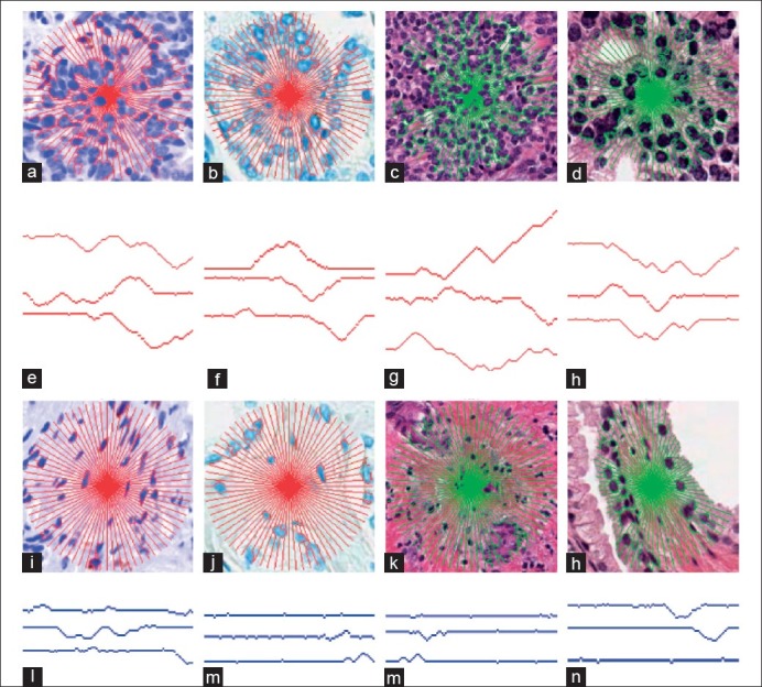
At every point of interest, the MS is determined as a series of radial paths extending outward in the direction of least resistance, navigating around obstructions. Each pixel can then be directly compared to other points of interest via a rotationally invariant quantitative feature descriptor, determined by the application of Fourier descriptors to the collection of these paths.

RESULTS

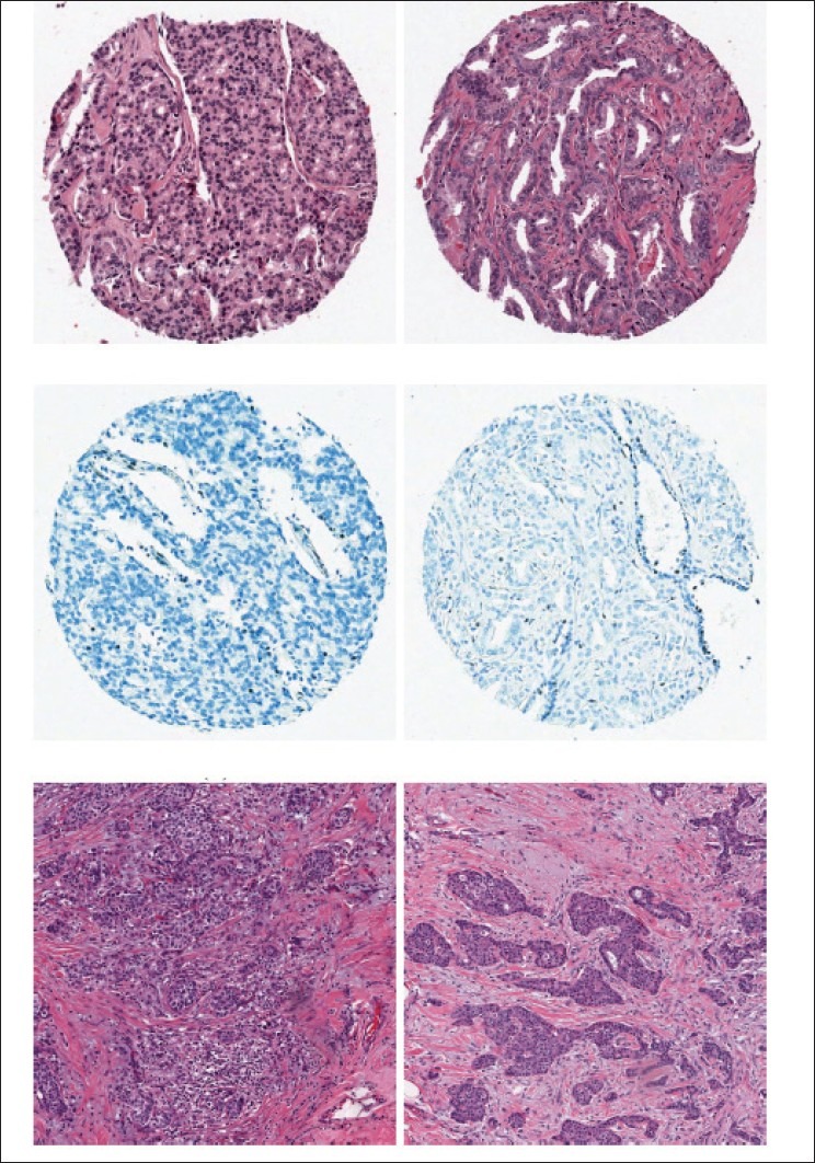
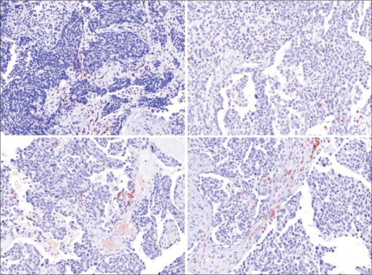
OUR GOAL IS TO DISTINGUISH TUMOR AND STROMAL TISSUE CLASSES IN THE CONTEXT OF FOUR DIFFERENT DIGITIZED PATHOLOGY DATASETS: prostate tissue microarrays (TMAs) stained with hematoxylin and eosin (HE) (44 images) and TMAs stained with only hematoxylin (H) (44 images), slide mounts of ovarian H (60 images), and HE breast cancer (51 images) histology images. Classification performance over 50 cross-validation runs using a Bayesian classifier produced mean areas under the curve of 0.88 ± 0.01 (prostate HE), 0.87 ± 0.02 (prostate H), 0.88 ± 0.01 (ovarian H), and 0.80 ± 0.01 (breast HE).

CONCLUSION

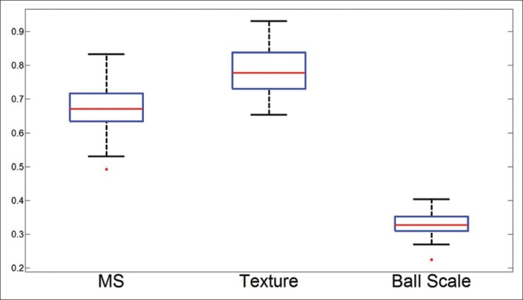
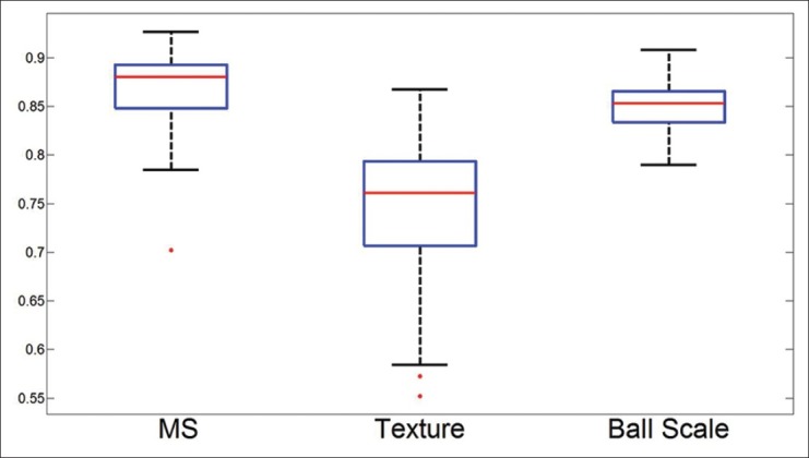
For each dataset listed in Table 3, we randomly selected 100 points per image, and using the procedure described in Experiment 1, we attempted to separate them as belonging to stroma or epithelium.

摘要

引言

引入局部尺度的概念是为了表征不同层次的图像细节,以便在执行局部图像处理任务的同时能够产生全局最优结果。在本文中,我们提出了一种新颖的局部自适应尺度定义——形态学尺度(MS)的方法框架,它与现有的局部尺度定义不同,因为它试图表征局部异质性而非局部同质性。

方法

在每个感兴趣的点上,MS被确定为一系列沿阻力最小方向向外延伸、绕过障碍物的径向路径。然后,通过将傅里叶描述符应用于这些路径的集合所确定的旋转不变定量特征描述符,可以将每个像素直接与其他感兴趣的点进行比较。

结果

我们的目标是在四个不同的数字化病理数据集的背景下区分肿瘤和基质组织类别:苏木精和伊红(HE)染色的前列腺组织微阵列(TMA)(44张图像)和仅苏木精(H)染色的TMA(44张图像)、卵巢H的载玻片(60张图像)以及HE乳腺癌(51张图像)组织学图像。使用贝叶斯分类器在50次交叉验证运行中的分类性能产生的曲线下平均面积为:0.88±0.01(前列腺HE)、0.87±0.02(前列腺H)、0.88±0.01(卵巢H)和0.80±0.01(乳腺HE)。

结论

对于表3中列出的每个数据集,我们每张图像随机选择100个点,并使用实验1中描述的程序,试图将它们区分为属于基质或上皮。

https://cdn.ncbi.nlm.nih.gov/pmc/blobs/3adb/3678744/6da24f89cdc3/JPI-4-8-g020.jpg
https://cdn.ncbi.nlm.nih.gov/pmc/blobs/3adb/3678744/6915dbb0c79b/JPI-4-8-g001.jpg
https://cdn.ncbi.nlm.nih.gov/pmc/blobs/3adb/3678744/eb79a9870817/JPI-4-8-g003.jpg
https://cdn.ncbi.nlm.nih.gov/pmc/blobs/3adb/3678744/a6b7047841af/JPI-4-8-g004.jpg
https://cdn.ncbi.nlm.nih.gov/pmc/blobs/3adb/3678744/8cd4fcb444cc/JPI-4-8-g007.jpg
https://cdn.ncbi.nlm.nih.gov/pmc/blobs/3adb/3678744/46716456c394/JPI-4-8-g008.jpg
https://cdn.ncbi.nlm.nih.gov/pmc/blobs/3adb/3678744/0128dba9de16/JPI-4-8-g009.jpg
https://cdn.ncbi.nlm.nih.gov/pmc/blobs/3adb/3678744/a4cc742b091f/JPI-4-8-g013.jpg
https://cdn.ncbi.nlm.nih.gov/pmc/blobs/3adb/3678744/ec84ab8b3cfa/JPI-4-8-g014.jpg
https://cdn.ncbi.nlm.nih.gov/pmc/blobs/3adb/3678744/8360593f1428/JPI-4-8-g015.jpg
https://cdn.ncbi.nlm.nih.gov/pmc/blobs/3adb/3678744/83ef20809ad6/JPI-4-8-g016.jpg
https://cdn.ncbi.nlm.nih.gov/pmc/blobs/3adb/3678744/d92ade65a0fa/JPI-4-8-g017.jpg
https://cdn.ncbi.nlm.nih.gov/pmc/blobs/3adb/3678744/84cc18a63f1c/JPI-4-8-g018.jpg
https://cdn.ncbi.nlm.nih.gov/pmc/blobs/3adb/3678744/6da24f89cdc3/JPI-4-8-g020.jpg

相似文献

1
Quantifying local heterogeneity via morphologic scale: Distinguishing tumoral from stromal regions.通过形态学尺度量化局部异质性:区分肿瘤区域和基质区域。
J Pathol Inform. 2013 Mar 30;4(Suppl):S8. doi: 10.4103/2153-3539.109865. Print 2013.
2
Machine learning approaches to analyze histological images of tissues from radical prostatectomies.用于分析前列腺癌根治术组织学图像的机器学习方法。
Comput Med Imaging Graph. 2015 Dec;46 Pt 2(Pt 2):197-208. doi: 10.1016/j.compmedimag.2015.08.002. Epub 2015 Aug 20.
3
Identification of tumor epithelium and stroma in tissue microarrays using texture analysis.利用纹理分析鉴定组织微阵列中的肿瘤上皮和基质。
Diagn Pathol. 2012 Mar 2;7:22. doi: 10.1186/1746-1596-7-22.
4
A new optical density granulometry-based descriptor for the classification of prostate histological images using shallow and deep Gaussian processes.一种基于新光学密度粒度分析的描述符,用于使用浅层和深层高斯过程对前列腺组织学图像进行分类。
Comput Methods Programs Biomed. 2019 Sep;178:303-317. doi: 10.1016/j.cmpb.2019.07.003. Epub 2019 Jul 4.
5
Content-based image retrieval of digitized histopathology in boosted spectrally embedded spaces.在增强的光谱嵌入空间中基于内容的数字化组织病理学图像检索
J Pathol Inform. 2015 Jun 29;6:41. doi: 10.4103/2153-3539.159441. eCollection 2015.
6
Semi-supervised training of deep convolutional neural networks with heterogeneous data and few local annotations: An experiment on prostate histopathology image classification.基于异构数据和少量局部标注的深度卷积神经网络的半监督学习:前列腺组织病理学图像分类实验。
Med Image Anal. 2021 Oct;73:102165. doi: 10.1016/j.media.2021.102165. Epub 2021 Jul 14.
7
Use of Deep Learning to Develop and Analyze Computational Hematoxylin and Eosin Staining of Prostate Core Biopsy Images for Tumor Diagnosis.使用深度学习开发和分析前列腺核心活检图像的计算苏木精和伊红染色,用于肿瘤诊断。
JAMA Netw Open. 2020 May 1;3(5):e205111. doi: 10.1001/jamanetworkopen.2020.5111.
8
An optimized color transformation for the analysis of digital images of hematoxylin & eosin stained slides.一种用于苏木精和伊红染色玻片数字图像分析的优化颜色转换方法。
J Pathol Inform. 2015 Jun 23;6:33. doi: 10.4103/2153-3539.158910. eCollection 2015.
9
New breast cancer prognostic factors identified by computer-aided image analysis of HE stained histopathology images.通过对苏木精-伊红(HE)染色的组织病理学图像进行计算机辅助图像分析确定的新的乳腺癌预后因素。
Sci Rep. 2015 May 29;5:10690. doi: 10.1038/srep10690.
10
A gamma-gaussian mixture model for detection of mitotic cells in breast cancer histopathology images.一种用于在乳腺癌组织病理学图像中检测有丝分裂细胞的伽马-高斯混合模型。
J Pathol Inform. 2013 May 30;4:11. doi: 10.4103/2153-3539.112696. Print 2013.

引用本文的文献

1
An alternative reference space for H&E color normalization.苏木精-伊红染色颜色标准化的另一种参考空间。
PLoS One. 2017 Mar 29;12(3):e0174489. doi: 10.1371/journal.pone.0174489. eCollection 2017.
2
A method for comparing intra-tumoural radioactivity uptake heterogeneity in preclinical positron emission tomography studies.一种在临床前正电子发射断层扫描研究中比较肿瘤内放射性摄取异质性的方法。
EJNMMI Phys. 2015 Dec;2(1):19. doi: 10.1186/s40658-015-0124-1. Epub 2015 Sep 8.
3
Beyond immune density: critical role of spatial heterogeneity in estrogen receptor-negative breast cancer.

本文引用的文献

1
Identification of tumor epithelium and stroma in tissue microarrays using texture analysis.利用纹理分析鉴定组织微阵列中的肿瘤上皮和基质。
Diagn Pathol. 2012 Mar 2;7:22. doi: 10.1186/1746-1596-7-22.
2
High-throughput biomarker segmentation on ovarian cancer tissue microarrays via hierarchical normalized cuts.基于层次归一化切割的卵巢癌组织微阵列高通量生物标志物分割。
IEEE Trans Biomed Eng. 2012 May;59(5):1240-52. doi: 10.1109/TBME.2011.2179546. Epub 2011 Dec 13.
3
Automatic tumor-stroma separation in fluorescence TMAs enables the quantitative high-throughput analysis of multiple cancer biomarkers.
超越免疫密度:空间异质性在雌激素受体阴性乳腺癌中的关键作用
Mod Pathol. 2015 Jun;28(6):766-77. doi: 10.1038/modpathol.2015.37. Epub 2015 Feb 27.
4
Novel genotype-phenotype associations in human cancers enabled by advanced molecular platforms and computational analysis of whole slide images.先进分子平台及全切片图像计算分析助力下的人类癌症新型基因型-表型关联
Lab Invest. 2015 Apr;95(4):366-76. doi: 10.1038/labinvest.2014.153. Epub 2015 Jan 19.
5
Mapping spatial heterogeneity in the tumor microenvironment: a new era for digital pathology.绘制肿瘤微环境中的空间异质性:数字病理学的新时代。
Lab Invest. 2015 Apr;95(4):377-84. doi: 10.1038/labinvest.2014.155. Epub 2015 Jan 19.
荧光 TMA 中的自动肿瘤-基质分离可实现多种癌症生物标志物的高通量定量分析。
PLoS One. 2011;6(12):e28048. doi: 10.1371/journal.pone.0028048. Epub 2011 Dec 2.
4
Systematic analysis of breast cancer morphology uncovers stromal features associated with survival.系统分析乳腺癌形态学揭示了与生存相关的基质特征。
Sci Transl Med. 2011 Nov 9;3(108):108ra113. doi: 10.1126/scitranslmed.3002564.
5
A high-throughput active contour scheme for segmentation of histopathological imagery.一种用于组织病理学图像分割的高通量主动轮廓方案。
Med Image Anal. 2011 Dec;15(6):851-62. doi: 10.1016/j.media.2011.04.002. Epub 2011 Apr 28.
6
Tensor classification of N-point correlation function features for histology tissue segmentation.用于组织学组织分割的 N 点相关函数特征的张量分类
Med Image Anal. 2009 Feb;13(1):156-66. doi: 10.1016/j.media.2008.06.020. Epub 2008 Jul 25.
7
Automated region of interest retrieval and classification using spectral analysis.利用光谱分析进行自动感兴趣区域提取和分类。
Diagn Pathol. 2008 Jul 15;3 Suppl 1(Suppl 1):S17. doi: 10.1186/1746-1596-3-S1-S17.
8
Locally adaptive perceptual image coding.局部自适应感知图像编码。
IEEE Trans Image Process. 2000;9(9):1472-83. doi: 10.1109/83.862622.
9
New methods of MR image intensity standardization via generalized scale.基于广义尺度的磁共振图像强度标准化新方法。
Med Phys. 2006 Sep;33(9):3426-34. doi: 10.1118/1.2335487.
10
Intraepithelial CD8+ tumor-infiltrating lymphocytes and a high CD8+/regulatory T cell ratio are associated with favorable prognosis in ovarian cancer.上皮内CD8 +肿瘤浸润淋巴细胞和高CD8 + /调节性T细胞比率与卵巢癌的良好预后相关。
Proc Natl Acad Sci U S A. 2005 Dec 20;102(51):18538-43. doi: 10.1073/pnas.0509182102. Epub 2005 Dec 12.