Laboratory of Rheumatology, GIGA Research, University of Liège, CHU Liège, Liège, Belgium.
PLoS One. 2013 Jun 12;8(6):e66769. doi: 10.1371/journal.pone.0066769. Print 2013.
To determine if serum amyloid A (A-SAA) could be detected in human osteoarthritic (OA) joints and further clarify if high A-SAA level in joints result from a local production or from a diffusion process from abnormally elevated plasma concentration. Regulatory mechanism of A-SAA expression and its pro-inflammatory properties were also investigated.
A-SAA levels in serum and synovial fluid of OA (n = 29) and rheumatoid arthritis (RA) (n = 27) patients were measured and compared to matched-healthy volunteers (HV) (n = 35). In vitro cell cultures were performed on primary joint cells provided from osteoarthritis patients. Regulatory mechanisms were studied using Western-blotting, ELISA and lentiviral transfections.
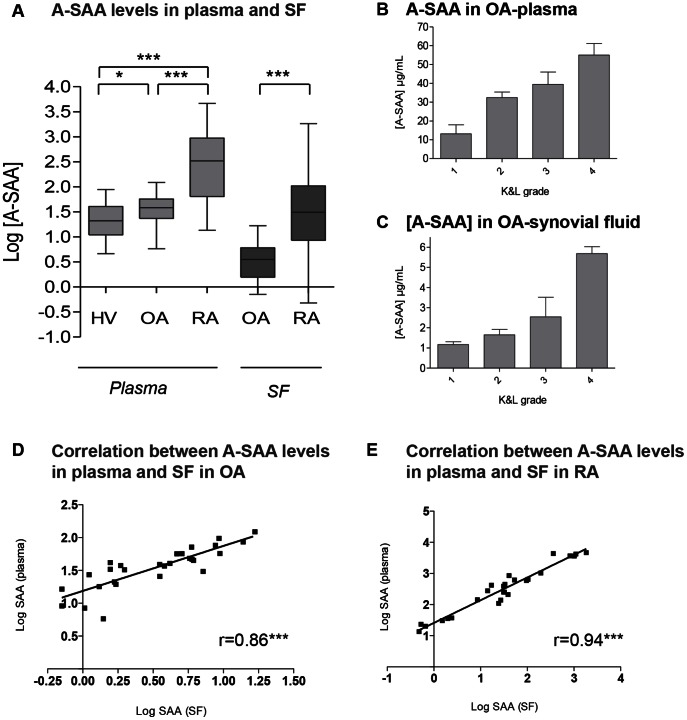
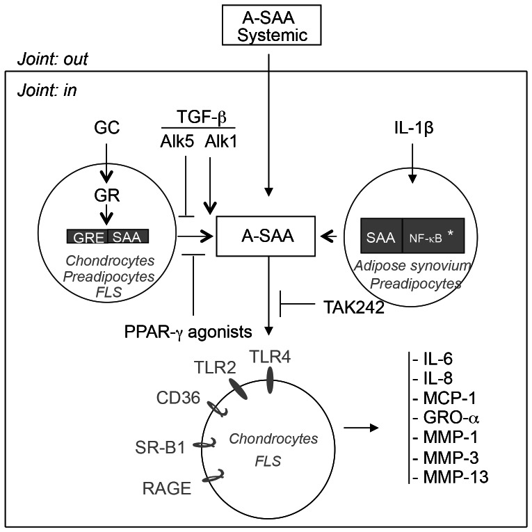
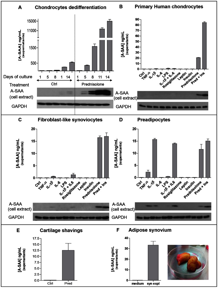
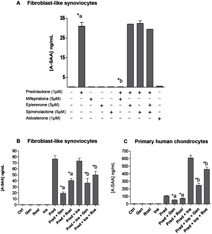
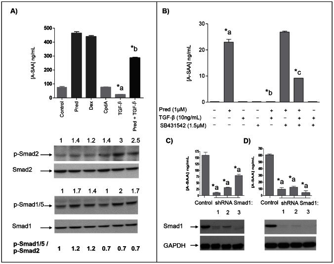
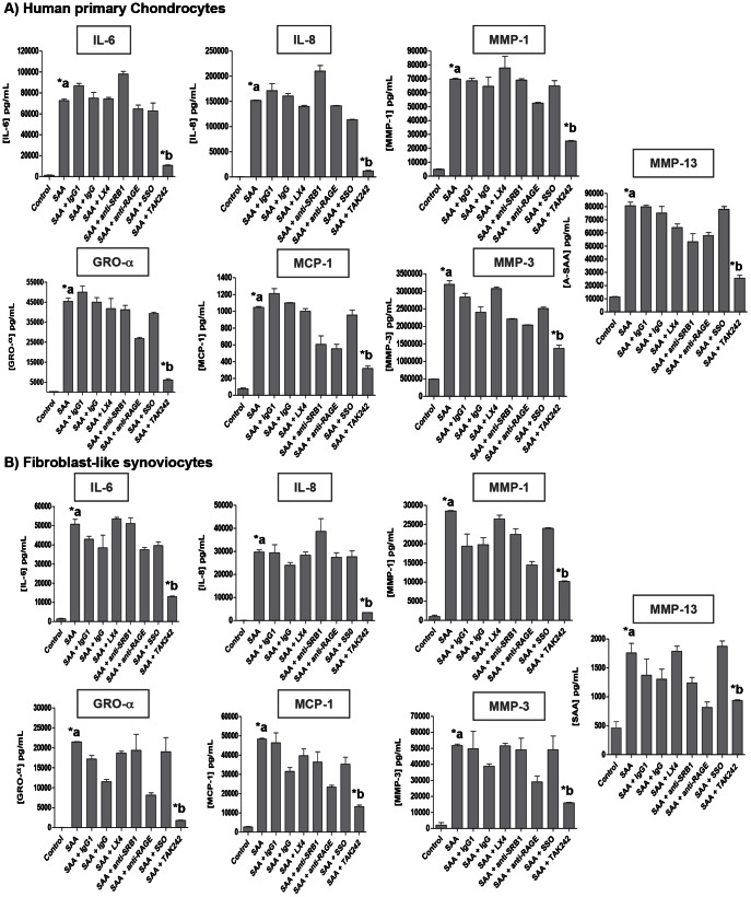
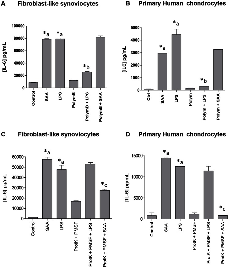
A-SAA was statistically increased in OA plasma patients compared to HV. Moreover, A-SAA level in OA plasma and synovial fluid increased with the Kellgren & Lauwrence grade. For all OA and RA patients, A-SAA plasma level was higher and highly correlated with its corresponding level in the synovial fluid, therefore supporting that A-SAA was mainly due to the passive diffusion process from blood into the joint cavity. However, A-SAA expression was also observed in vitro under corticosteroid treatment and/or under IL-1beta stimuli. A-SAA expression was down-regulated by PPAR-γ agonists (genistein and rosiglitazone) and up-regulated by TGF-β1 through Alk1 (Smad1/5) pathway. RhSAA induced proinflammatory cytokines (IL-6, IL-8, GRO-α and MCP-1) and metalloproteinases (MMP-1, MMP-3 and MMP-13) expression in FLS and chondrocytes, which expression was downregulated by TAK242, a specific TLR4 inhibitor.
Systemic or local A-SAA expression inside OA joint cavity may play a key role in inflammatory process seen in osteoarthritis, which could be counteracted by TLR4 inhibition.
确定血清淀粉样蛋白 A(A-SAA)是否可在人类骨关节炎(OA)关节中检测到,并进一步阐明关节中高 A-SAA 水平是源于局部产生还是源于异常升高的血浆浓度的扩散过程。还研究了 A-SAA 表达的调节机制及其促炎特性。
测量了 29 例 OA 患者和 27 例类风湿关节炎(RA)患者的血清和滑液中 A-SAA 水平,并与相匹配的健康志愿者(HV)(n=35)进行了比较。对来自骨关节炎患者的原代关节细胞进行了体外细胞培养。使用 Western 印迹、ELISA 和慢病毒转染研究了调节机制。
与 HV 相比,OA 患者的血浆 A-SAA 统计学上增加。此外,OA 患者的血浆和滑液中的 A-SAA 水平随着 Kellgren & Lauwrence 分级而增加。对于所有 OA 和 RA 患者,A-SAA 的血浆水平更高,并且与相应的滑液水平高度相关,因此支持 A-SAA 主要是由于从血液到关节腔的被动扩散过程。然而,在皮质类固醇治疗和/或在 IL-1β刺激下,也在体外观察到 A-SAA 的表达。PPAR-γ 激动剂(染料木黄酮和罗格列酮)可下调 A-SAA 表达,TGF-β1 通过 Alk1(Smad1/5)途径可上调 A-SAA 表达。RhSAA 在 FLS 和软骨细胞中诱导促炎细胞因子(IL-6、IL-8、GRO-α 和 MCP-1)和金属蛋白酶(MMP-1、MMP-3 和 MMP-13)的表达,TAK242(一种特异性 TLR4 抑制剂)可下调其表达。
OA 关节腔内的全身性或局部 A-SAA 表达可能在骨关节炎中所见的炎症过程中起关键作用,TLR4 抑制可能对此起拮抗作用。