Biological Sciences Division, Pacific Northwest National Laboratory, Richland, Washington, United States of America.
PLoS One. 2013 Jun 14;8(6):e65874. doi: 10.1371/journal.pone.0065874. Print 2013.
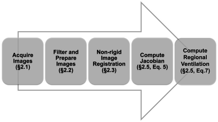
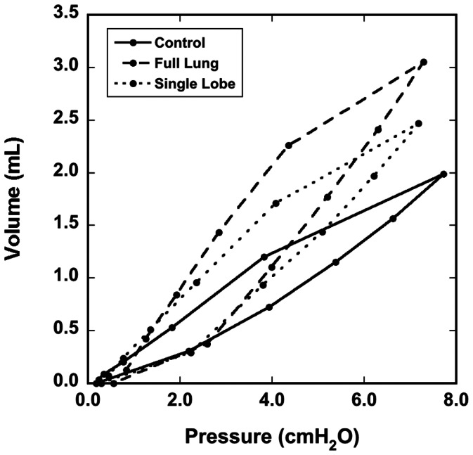
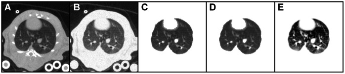
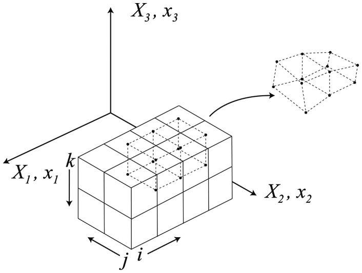
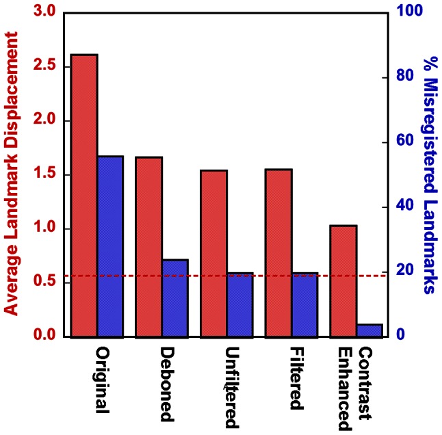
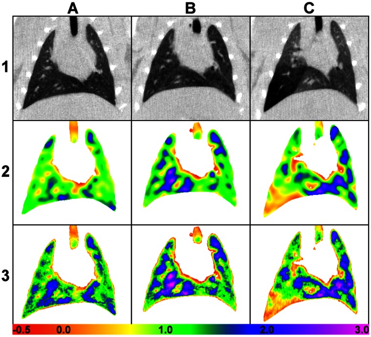
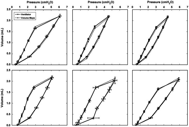
Changes in the shape of the lung during breathing determine the movement of airways and alveoli, and thus impact airflow dynamics. Modeling airflow dynamics in health and disease is a key goal for predictive multiscale models of respiration. Past efforts to model changes in lung shape during breathing have measured shape at multiple breath-holds. However, breath-holds do not capture hysteretic differences between inspiration and expiration resulting from the additional energy required for inspiration. Alternatively, imaging dynamically--without breath-holds--allows measurement of hysteretic differences. In this study, we acquire multiple micro-CT images per breath (4DCT) in live rats, and from these images we develop, for the first time, dynamic volume maps. These maps show changes in local volume across the entire lung throughout the breathing cycle and accurately predict the global pressure-volume (PV) hysteresis. Male Sprague-Dawley rats were given either a full- or partial-lung dose of elastase or saline as a control. After three weeks, 4DCT images of the mechanically ventilated rats under anesthesia were acquired dynamically over the breathing cycle (11 time points, ≤100 ms temporal resolution, 8 cmH2O peak pressure). Non-rigid image registration was applied to determine the deformation gradient--a numerical description of changes to lung shape--at each time point. The registration accuracy was evaluated by landmark identification. Of 67 landmarks, one was determined misregistered by all three observers, and 11 were determined misregistered by two observers. Volume change maps were calculated on a voxel-by-voxel basis at all time points using both the Jacobian of the deformation gradient and the inhaled air fraction. The calculated lung PV hysteresis agrees with pressure-volume curves measured by the ventilator. Volume maps in diseased rats show increased compliance and ventilation heterogeneity. Future predictive multiscale models of rodent respiration may leverage such volume maps as boundary conditions.
在呼吸过程中,肺部形状的变化决定了气道和肺泡的运动,从而影响气流动力学。在健康和疾病中对气流动力学进行建模是呼吸多尺度预测模型的一个关键目标。过去在呼吸过程中对肺部形状变化进行建模的努力已经在多次呼吸暂停中测量了形状。然而,呼吸暂停并没有捕捉到由于吸气所需的额外能量而导致的吸气和呼气之间的滞后差异。相比之下,动态成像(无需呼吸暂停)可以测量滞后差异。在这项研究中,我们在活老鼠身上每呼吸一次获取多个微 CT 图像(4DCT),并从这些图像中首次开发了动态体积图。这些图谱显示了整个肺部在整个呼吸周期中局部体积的变化,并准确预测了全局压力-容积(PV)滞后。雄性 Sprague-Dawley 大鼠接受弹性蛋白酶全肺或半肺剂量或生理盐水作为对照。三周后,对机械通气的麻醉大鼠在呼吸周期内(11 个时间点,≤100ms 时间分辨率,8cmH2O 峰值压力)动态获取 4DCT 图像。应用非刚性图像配准来确定变形梯度——肺部形状变化的数值描述——在每个时间点。通过标志点识别评估配准精度。在 67 个标志点中,有一个标志点被所有三个观察者确定为配准错误,有 11 个标志点被两个观察者确定为配准错误。在所有时间点上,使用变形梯度的雅可比和吸入空气分数在体素基础上计算体积变化图。计算得到的肺 PV 滞后与通气机测量的压力-容积曲线一致。患病大鼠的体积图显示顺应性增加和通气异质性增加。未来的啮齿动物呼吸多尺度预测模型可能会利用这些体积图作为边界条件。