BEEgroup, Biocentre, University of Würzburg, Würzburg, Germany.
PLoS One. 2013 Jun 17;8(6):e66415. doi: 10.1371/journal.pone.0066415. Print 2013.
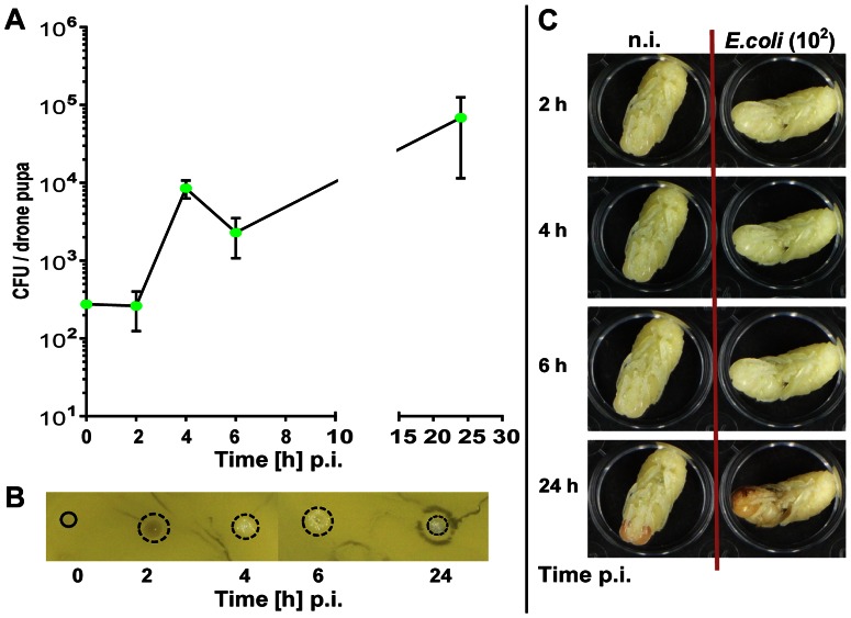
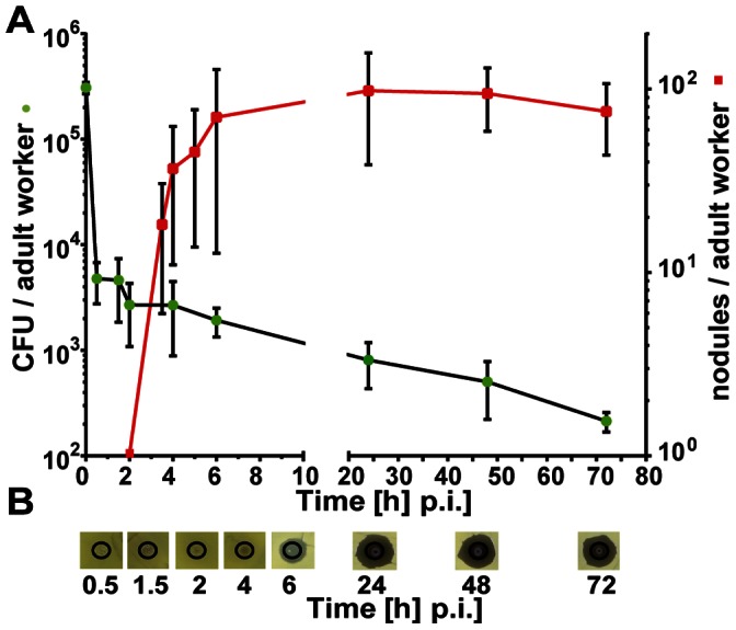
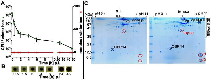
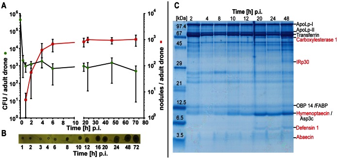
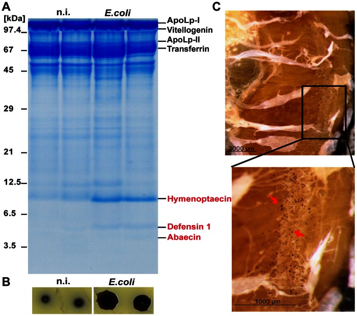
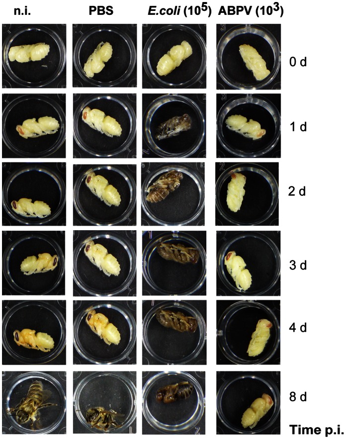
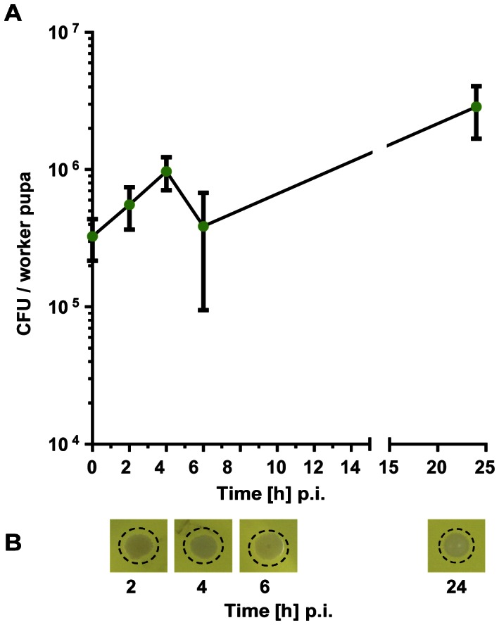
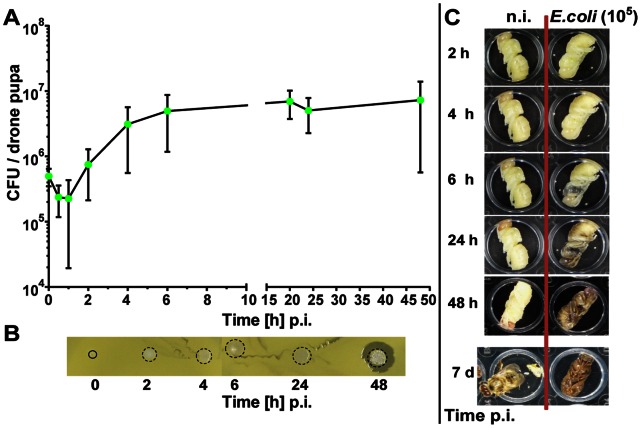
The development of all honey bee castes proceeds through three different life stages all of which encounter microbial infections to a various extent. We have examined the immune strength of honey bees across all developmental stages with emphasis on the temporal expression of cellular and humoral immune responses upon artificial challenge with viable Escherichia coli bacteria. We employed a broad array of methods to investigate defence strategies of infected individuals: (a) fate of bacteria in the haemocoel; (b) nodule formation and (c) induction of antimicrobial peptides (AMPs). Newly emerged adult worker bees and drones were able to activate efficiently all examined immune reactions. The number of viable bacteria circulating in the haemocoel of infected bees declined rapidly by more than two orders of magnitude within the first 4-6 h post-injection (p.i.), coinciding with the occurrence of melanised nodules. Antimicrobial activity, on the other hand, became detectable only after the initial bacterial clearance. These two temporal patterns of defence reactions very likely represent the constitutive cellular and the induced humoral immune response. A unique feature of honey bees is that a fraction of worker bees survives the winter season in a cluster mostly engaged in thermoregulation. We show here that the overall immune strength of winter bees matches that of young summer bees although nodulation reactions are not initiated at all. As expected, high doses of injected viable E.coli bacteria caused no mortality in larvae or adults of each age. However, drone and worker pupae succumbed to challenge with E.coli even at low doses, accompanied by a premature darkening of the pupal body. In contrast to larvae and adults, we observed no fast clearance of viable bacteria and no induction of AMPs but a rapid proliferation of E.coli bacteria in the haemocoel of bee pupae ultimately leading to their death.
所有蜜蜂蜂型的发育都经过三个不同的生命阶段,这些阶段都在不同程度上遭遇微生物感染。我们检查了蜜蜂在所有发育阶段的免疫强度,重点研究了在受到活大肠杆菌人工挑战时细胞和体液免疫反应的时间表达。我们采用了广泛的方法来研究受感染个体的防御策略:(a)血腔中细菌的命运;(b)结节的形成和(c)抗菌肽(AMPs)的诱导。新出现的成年工蜂和雄蜂能够有效地激活所有检查的免疫反应。感染蜜蜂血腔中循环的活菌数量在注射后 4-6 小时内迅速下降两个数量级以上,这与黑化结节的发生相吻合。另一方面,抗菌活性仅在初始细菌清除后才变得可检测。这两种防御反应的时间模式很可能代表了组成性细胞免疫和诱导性体液免疫反应。蜜蜂的一个独特特征是,一部分工蜂在一个主要从事体温调节的群体中幸存下来度过冬季。我们在这里表明,冬季蜜蜂的整体免疫强度与年轻的夏季蜜蜂相当,尽管结节反应根本没有启动。正如预期的那样,高剂量的注射活大肠杆菌细菌在每个年龄段的幼虫或成虫中都不会引起死亡。然而,雄蜂和工蜂蛹在低剂量的大肠杆菌挑战下就会死亡,同时蛹体迅速变黑。与幼虫和成虫不同,我们观察到活细菌没有快速清除,也没有诱导 AMPs,但在蜜蜂蛹的血腔中大肠杆菌细菌迅速增殖,最终导致它们死亡。