Department of Otorhinolaryngology, Dankook University College of Medicine, Cheonan, Korea. ; Beckman Laser Institute-Korea, Dankook University, Cheonan, Korea.
Clin Exp Otorhinolaryngol. 2013 Jun;6(2):82-9. doi: 10.3342/ceo.2013.6.2.82. Epub 2013 Jun 14.
Cartilage reshaping by laser irradiation is used to correct septal and auricular cartilage deformities. Chondrocyte viability following laser irradiation and reshaping has been well established. However, the regeneration process of chondrocyte after laser irradiation has not been revealed yet. The aims of this study were to determine the mechanism of cartilaginous thermal injury and the regenerative process of damaged cartilage following laser irradiation.
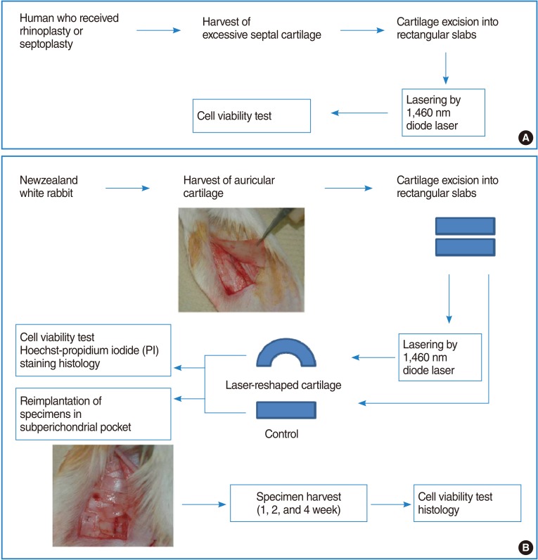
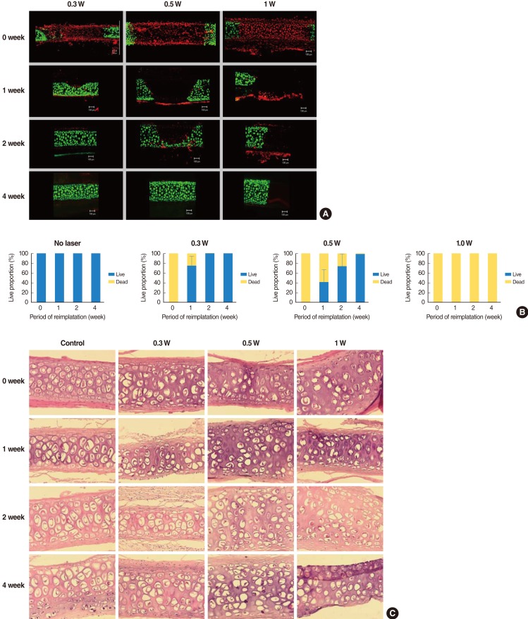
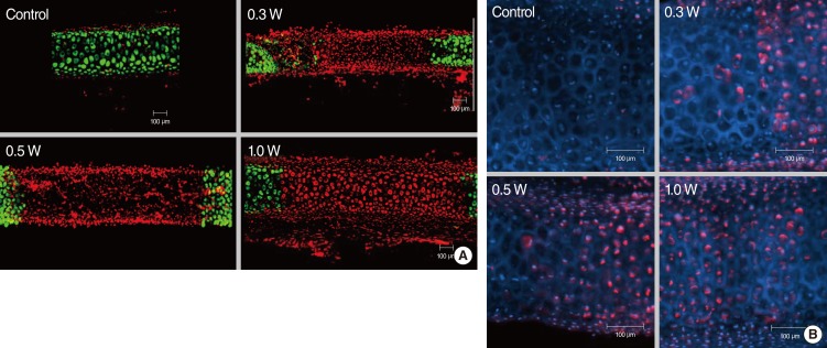
Laser irradiation was performed on human septal cartilage and rabbit auricular cartilage using a 1,460-nm diode laser. We observed change in the shape of cartilage and evaluated the extent of cartilage injury using live/dead cell assay via confocal microscopy. Hoechst and propidium iodide (PI) staining was used to evaluate the mechanism of chondrocyte injury after laser irradiation. To evaluate the regeneration of cartilage, laser irradiated cartilages were reimplanted into a subperichondrial pocket and were harvested at 1, 2, and 4 weeks after reimplantation for viability assessment and histologic examination.
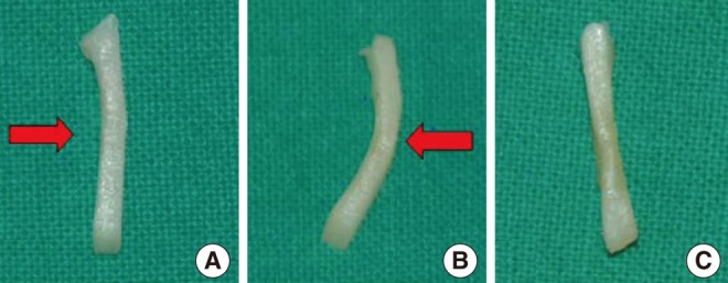
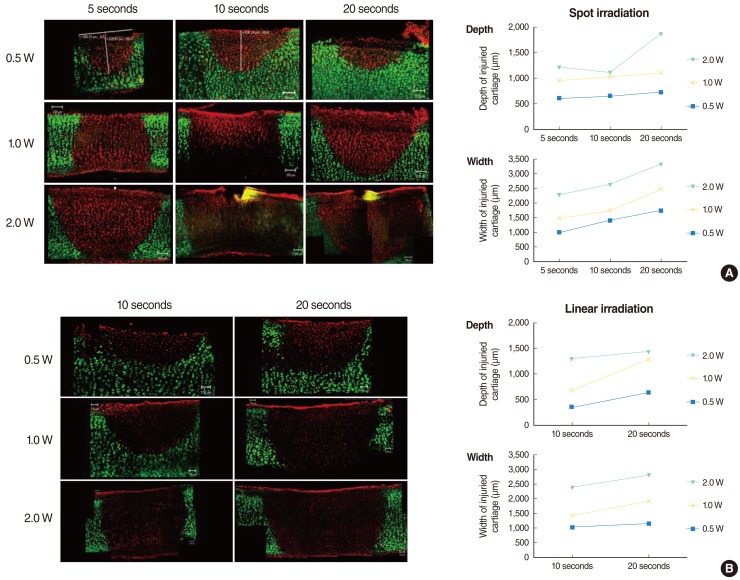
Laser irradiation using a 1,460-nm diode laser produced a marked shape change in both human septal and rabbit auricular cartilages. Thermal damage on cartilage was correlated with the exposure time and the laser power. Hoechst and PI staining showed that chondrocyte death by laser irradiation was due to mainly necrosis, rather than apoptosis. In lower power treatment group (0.3 W and 0.5 W), all the chondrocytes regenerated within 4 weeks, however, in 1 W treatment group, chondrocytes could not regenerate until 4 weeks.
Reshaping of cartilage using 1,460 nm diode laser was attained concurrently with the thermal injury to the chondrocytes. The extent of thermal damage on chondrocytes was dependent on the exposure time and the laser power and the damaged chondrocytes irradiated with lower level of laser power could be regenerated after reimplantation into subperichondrial pocket.
通过激光照射重塑软骨,用于矫正鼻中隔和耳廓软骨畸形。激光照射后软骨细胞的活力已得到充分证实。然而,激光照射后软骨细胞的再生过程尚未揭示。本研究旨在确定软骨热损伤的机制以及激光照射后软骨损伤的再生过程。
采用 1460nm 二极管激光对人鼻中隔软骨和兔耳廓软骨进行激光照射。我们通过共聚焦显微镜观察软骨形状的变化,并通过活/死细胞检测评估软骨损伤的程度。Hoechst 和碘化丙啶(PI)染色用于评估激光照射后软骨细胞损伤的机制。为了评估软骨的再生,将激光照射的软骨重新植入软骨下囊中,并在重新植入后 1、2 和 4 周进行活组织评估和组织学检查。
使用 1460nm 二极管激光照射产生了人鼻中隔和兔耳廓软骨的明显形状变化。软骨的热损伤与暴露时间和激光功率有关。Hoechst 和 PI 染色表明,激光照射导致的软骨细胞死亡主要是坏死,而不是凋亡。在较低功率处理组(0.3W 和 0.5W)中,所有软骨细胞在 4 周内再生,然而,在 1W 处理组中,软骨细胞直到 4 周后才能够再生。
使用 1460nm 二极管激光重塑软骨的同时也对软骨细胞造成了热损伤。软骨细胞的热损伤程度取决于暴露时间和激光功率,较低水平激光照射的损伤软骨在重新植入软骨下囊中后可以再生。