Ribeiro Rejane G, Costa Ana Paula A, Bragato Nathália, Fonseca Angela M, Duque Juan C M, Prado Tales D, Silva Andrea C R, Borges Naida C
BMC Vet Res. 2013 Jun 23;9:124. doi: 10.1186/1746-6148-9-124.
The use of ultrasound in veterinary medicine is widespread as a diagnostic supplement in the clinical routine of small animals, but there are few reports in wild animals. The objective of this study was to describe the anatomy, topography and abdominal sonographic features of coatis.
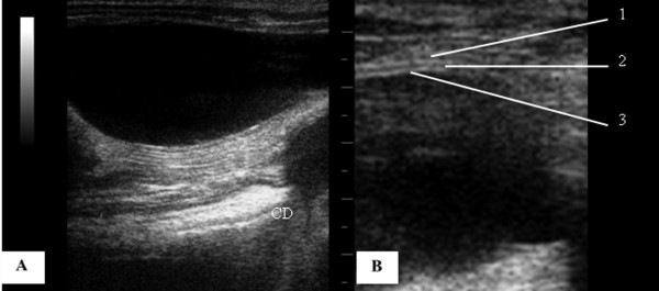
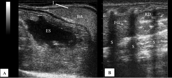
The urinary bladder wall measured 0.11 ± 0.03 cm. The symmetrical kidneys were in the left and right cranial quadrant of the abdomen and the cortical, medullary and renal pelvis regions were recognized and in all sections. The medullary rim sign was visualized in the left kidney of two coatis. The liver had homogeneous texture and was in the cranial abdomen under the rib cage. The gallbladder, rounded and filled with anechoic content was visualized in all coatis, to the right of the midline. The spleen was identified in the left cranial abdomen following the greater curvature of the stomach. The parenchyma was homogeneous and hyperechogenic compared to the liver and kidney cortex. The stomach was in the cranial abdomen, limited cranially by the liver and caudo-laterally by the spleen. The left adrenal glands of five coatis were seen in the cranial pole of the left kidney showing hypoechogenic parenchyma without distinction of cortex and medulla. The pancreas was visualized in only two coatis. The left ovary (0.92 cm x 0.56 cm) was visualized on a single coati in the caudal pole of the kidney. The uterus, right adrenal, right ovary and intestines were not visualized.
Ultrasound examination of the abdomen of coatis may be accomplished by following the recommendations for dogs and cats. It is possible to evaluate the anatomical and topographical relationships of the abdominal organs together with the knowledge of the peculiarities of parenchymal echogenicity and echotexture of the viscera.
超声在兽医学中作为小动物临床常规诊断的辅助手段应用广泛,但在野生动物中的报道较少。本研究的目的是描述南美浣熊的解剖结构、位置及腹部超声特征。
膀胱壁厚度为0.11±0.03厘米。双侧肾脏位于腹部左右上象限,皮质、髓质和肾盂区域在所有切面均可识别。两只南美浣熊的左肾可见髓质边缘征。肝脏质地均匀,位于胸腔下方的上腹部。所有南美浣熊均在中线右侧可见圆形且充满无回声内容物的胆囊。脾脏位于左上腹,沿胃大弯分布。与肝脏和肾皮质相比,脾实质均匀且回声增强。胃位于上腹部,上方由肝脏界定,后外侧由脾脏界定。五只南美浣熊的左肾上腺位于左肾的上极,显示为低回声实质,无法区分皮质和髓质。仅在两只南美浣熊中观察到胰腺。在一只南美浣熊的肾下极可见左侧卵巢(0.92厘米×0.56厘米)。未观察到子宫、右肾上腺、右卵巢和肠道。
按照犬猫的检查建议可对南美浣熊腹部进行超声检查。结合内脏实质回声特性和回声纹理特点的知识,有可能评估腹部器官的解剖和位置关系。