Hashmani Khurram, Branch Matthew James, Sidney Laura Elizabeth, Dhillon Permesh Singh, Verma Megha, McIntosh Owen Douglas, Hopkinson Andrew, Dua Harminder Singh
Stem Cell Res Ther. 2013 Jun 24;4(3):75. doi: 10.1186/scrt226.
The corneal stroma is being increasingly recognized as a repository for stem cells. Like the limbal and endothelial niches, stromal stem cells often reside in the peripheral cornea and limbus. These peripheral and limbal corneal stromal cells (PLCSCs) are known to produce mesenchymal stem cells in vitro. Recently, a common corneal stromal and epithelial progenitor was hinted at. This study aims to examine the stem cell potential of corneal stromal cells and to investigate their epithelial transdifferentiation ability.
PLCSCs were grown in traditional Dulbecco modified Eagle medium (DMEM)-based keratocyte culture medium and an M199-based medium and analyzed for a profile of cell-surface markers by using flow cytometry and differentiated into mesenchymal phenotypes analyzed with quantitative polymerase chain reaction (qPCR) and histologic staining. PLCSCs in M199 were subsequently divided into subpopulations based on CD34 and CD105 expression by using fluorescence- activated cell sorting (FACS). Subpopulations were characterized by marker profile and mesenchymal differentiation ability. Both whole PLCSCs and subpopulations were also cultured for epithelial transdifferentiation.
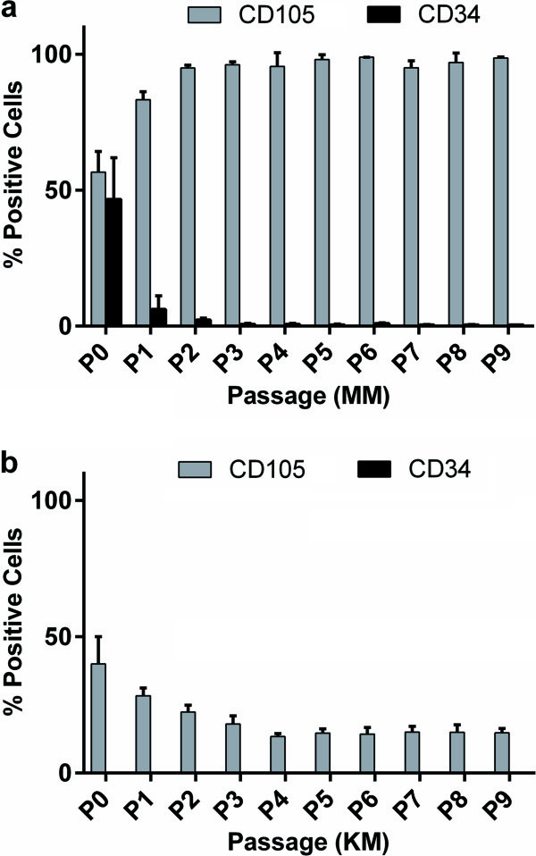
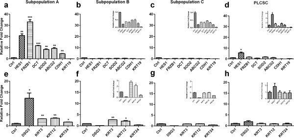
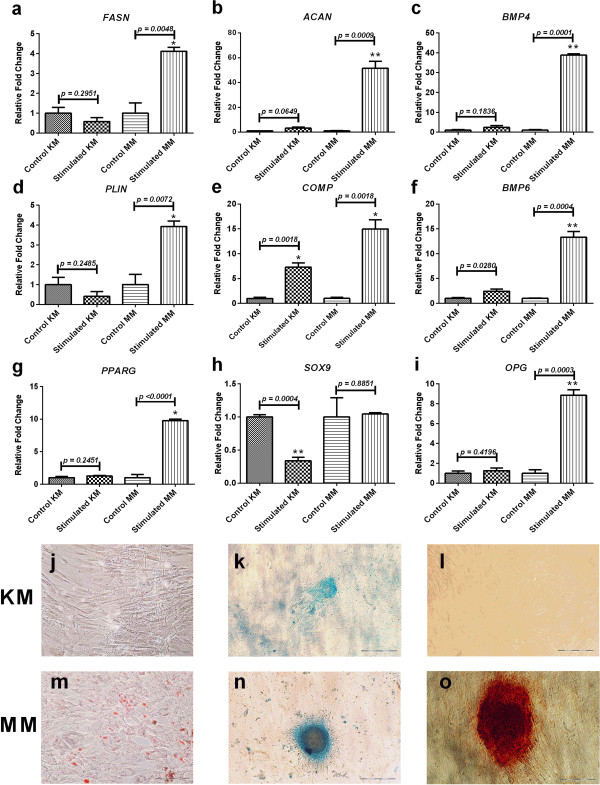
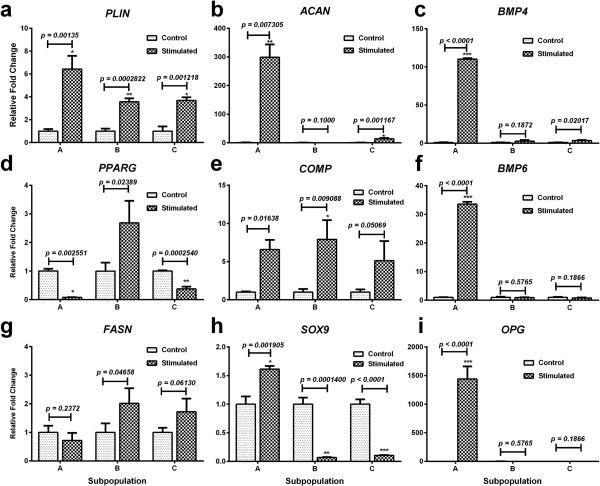
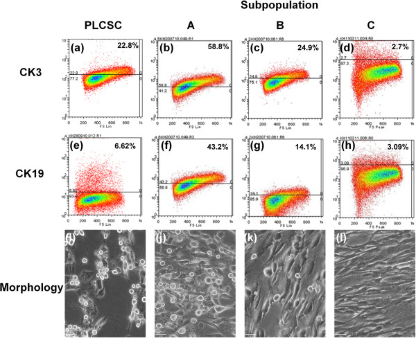
Cells cultured in M199 demonstrated a more stem-like cell-surface marker profile, and the keratocyte marker CD34 was retained for several passages but absent in cells cultured in DMEM. Cells cultured in M199 also exhibited a greater mesenchymal differentiation potential, compared with DMEM. PLCSCs could be divided into CD34(+)CD105(+), CD34-CD105(+), and CD34-CD105- subpopulations, of which CD34(+)CD105(+) cells were the most stemlike with regard to marker expression and mesenchymal differentiation potential. Subpopulations of PLCSCs exhibited differing abilities to transdifferentiate into epithelial phenotypes. Cells that were initially CD34(+)CD105(+) showed the greatest differentiation potential, producing CK3(+) and CK19(+) cells, and expressed a range of both epithelial progenitor (HES1, FRZB1, DCT, SOD2, ABCG2, CDH1, KRT19) and terminally differentiated (DSG3, KRT3, KRT12, KRT24) genes.
Culture medium has a significant effect on the phenotype and differentiation capacity of PLCSCs. The stroma contains a heterogeneous cell population in which we have identified CD34(+) cells as a stem cell population with a capacity for mesenchymal and epithelial differentiation.
角膜基质越来越被认为是干细胞的储存库。与角膜缘和内皮微环境一样,基质干细胞通常位于周边角膜和角膜缘。已知这些周边和角膜缘角膜基质细胞(PLCSCs)在体外可产生间充质干细胞。最近,有人暗示存在一种常见的角膜基质和上皮祖细胞。本研究旨在检测角膜基质细胞的干细胞潜能,并研究其上皮转分化能力。
将PLCSCs培养于传统的基于杜尔贝科改良伊格尔培养基(DMEM)的角膜细胞培养基和基于M199的培养基中,通过流式细胞术分析细胞表面标志物谱,并通过定量聚合酶链反应(qPCR)和组织学染色分析分化为间充质表型的情况。随后,利用荧光激活细胞分选(FACS)根据CD34和CD105表达将M199中的PLCSCs分为亚群。通过标志物谱和间充质分化能力对亚群进行表征。对整个PLCSCs及其亚群也进行上皮转分化培养。
在M199中培养的细胞表现出更具干细胞样的细胞表面标志物谱,角膜细胞标志物CD34在传代培养数代后仍保留,但在DMEM中培养的细胞中不存在。与DMEM相比,在M199中培养的细胞也表现出更大的间充质分化潜能。PLCSCs可分为CD34(+)CD105(+)、CD34-CD105(+)和CD34-CD105-亚群,其中CD34(+)CD105(+)细胞在标志物表达和间充质分化潜能方面最具干细胞样特征。PLCSCs亚群表现出不同的上皮转分化能力。最初为CD34(+)CD105(+)的细胞表现出最大的分化潜能,产生CK3(+)和CK19(+)细胞,并表达一系列上皮祖细胞(HES1、FRZB1、DCT、SOD2、ABCG2、CDH1、KRT19)和终末分化(DSG3、KRT3、KRT12、KRT24)基因。
培养基对PLCSCs的表型和分化能力有显著影响。基质包含异质性细胞群体,我们已确定CD34(+)细胞为具有间充质和上皮分化能力的干细胞群体。