Laboratory of Brain Research, Department of Neuroscience, Karolinska Institute Solna, Sweden.
Front Syst Neurosci. 2013 Jun 25;7:23. doi: 10.3389/fnsys.2013.00023. eCollection 2013.
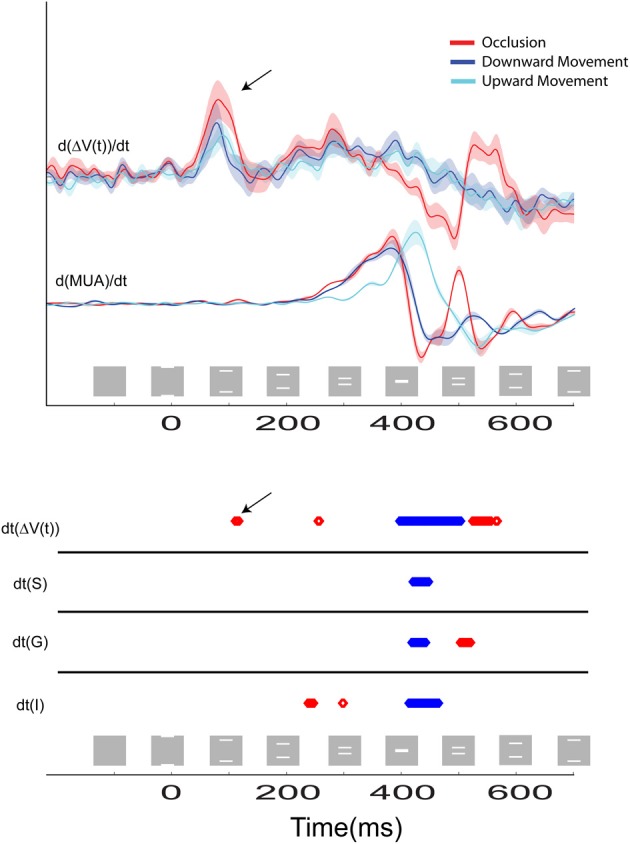
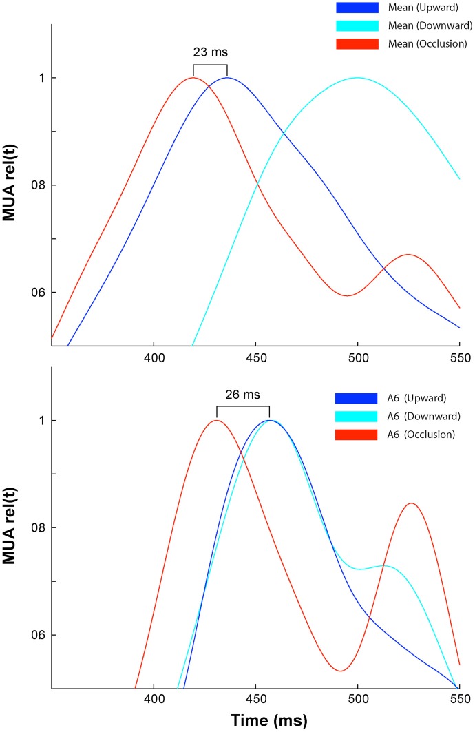
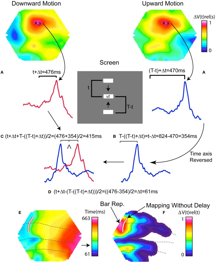
It is not known how visual cortical neurons react to several moving objects and how their firing to the motion of one object is affected by neurons firing to another moving object. Here we combine imaging of voltage sensitive dye (VSD) signals, reflecting the population membrane potential from ferret visual areas 17, 18, 19, and 21, with laminar recordings of multiunit activity, (MUA), when two bars moved toward each other in the visual field, occluded one another, and continued on in opposite directions. Two zones of peak MUA, mapping the bars' motion, moved toward each other along the area 17/18 border, which in the ferret maps the vertical meridian of the field of view. This was reflected also in the VSD signal, at both the 17/18 border as well as at the 19/21 border with a short delay. After some 125 ms at the area 19/21 border, the VSD signal increased and became elongated in the direction of motion in front of both of the moving representations. This was directly followed by the phase of the signal reversing and travelling back from the 19/21 border toward the 17/18 border, seemingly without respect for retinotopic boundaries, where it arrived at 150 ms after stimulus onset. At this point the VSD signal in front of the moving bar representations along the 17/18 border also increased and became elongated in the direction of object motion; the signal now being the linear sum of what has been observed in response to single moving bars. When the neuronal populations representing the bars were some 600 μm apart on the cortex, the dye signal and laminar MUA decreased strongly, with the MUA scaling to that of a single bar during occlusion. Despite a short rebound of the dye signal and MUA, the MUA after the occlusion was significantly depressed. The interactions between the neuronal populations mapping the bars' position, and the neurons in between these populations were, apart from 19/21 to 17/18 interaction, mainly lateral-horizontal; first excitatory and inducing firing at the site of future occlusion, then inhibitory just prior to occlusion. After occlusion the neurons that had fired already to the first bar showed delayed and prolonged inhibition in response to the second bar. Thus, the interactions that were particular to the occlusion condition in these experiments were local and inhibitory at short cortical range, and delayed and inhibitory after the occlusion when the bars moved further apart.
目前尚不清楚视觉皮层神经元如何对多个运动物体做出反应,以及一个物体的运动引起的神经元放电活动如何受到另一个运动物体的影响。在这里,我们结合电压敏感染料(VSD)信号成像,反映来自雪貂视觉区域 17、18、19 和 21 的群体膜电位,以及多单位活动(MUA)的分层记录,当两个棒状物在视野中彼此相向移动、相互遮挡并继续向相反方向移动时。两个 MUA 峰值区域映射棒状物的运动,沿着区域 17/18 边界相互靠近,在雪貂中,该区域映射视野的垂直子午线。这也反映在 VSD 信号中,在 17/18 边界以及 19/21 边界都有一个短暂的延迟。在区域 19/21 边界大约 125 毫秒后,VSD 信号增加,并在两个运动表示物前方的运动方向上拉长。紧随其后的是信号相位反转,从 19/21 边界向 17/18 边界移动,似乎不考虑视敏度边界,在刺激开始后 150 毫秒到达该边界。此时,在 17/18 边界处的运动棒状代表物前方的 VSD 信号也增加并沿物体运动方向拉长;该信号现在是对单个运动棒状刺激的反应的线性总和。当皮层上代表棒状物的神经元群体相隔约 600μm 时,染料信号和分层 MUA 强烈减弱,MUA 在遮挡时缩小为单个棒状物的 MUA。尽管染料信号和 MUA 有短暂的反弹,但遮挡后的 MUA 明显受到抑制。映射棒状位置的神经元群体之间以及这些群体之间的神经元之间的相互作用,除了 19/21 到 17/18 的相互作用外,主要是横向的;首先在未来遮挡处兴奋诱导放电,然后在遮挡前抑制。遮挡后,已经对第一根棒状刺激做出反应的神经元对第二根棒状刺激表现出延迟和持续的抑制。因此,在这些实验中,特定于遮挡条件的相互作用在短距离皮层内是局部和抑制性的,并且在棒状物进一步分开后,在遮挡后是延迟和抑制性的。