Department of Developmental Dentistry, Dental School, The University of Texas Health Science Center at San Antonio, 7703 Floyd Curl Drive, San Antonio, TX, 78229-3900, USA.
In Vitro Cell Dev Biol Anim. 2013 Sep;49(8):626-37. doi: 10.1007/s11626-013-9641-1. Epub 2013 Jun 29.
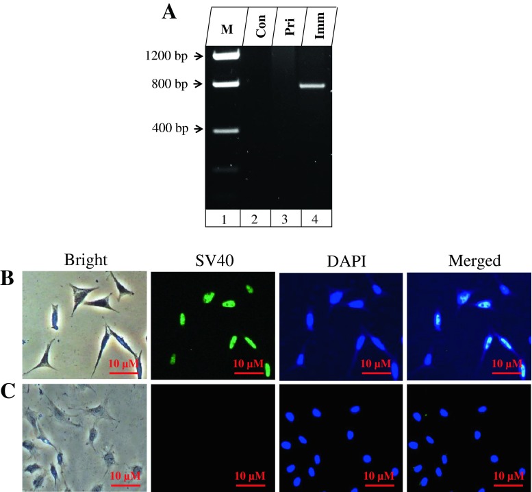
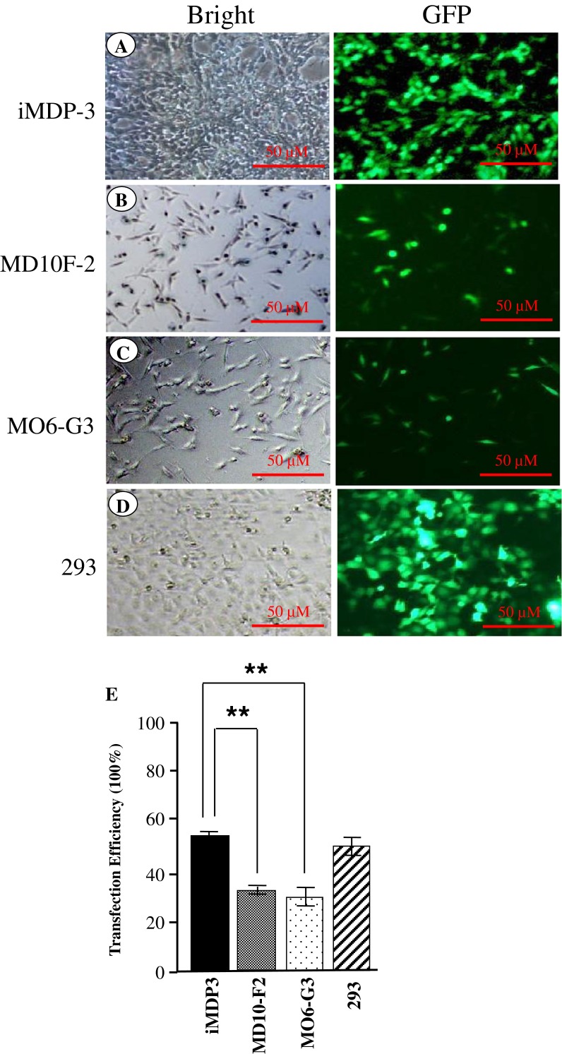
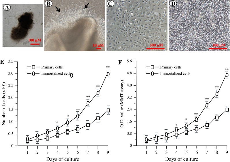
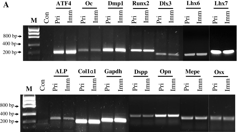
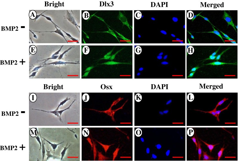
Odontogenesis is the result of the reciprocal interactions between epithelial-mesenchymal cells leading to terminally differentiated odontoblasts. This process from dental papilla mesenchymal cells to odontoblasts is regulated by a complex signaling pathway. When isolated from the developing tooth germs, odontoblasts quickly lose their potential to maintain the odontoblast-specific phenotype. Therefore, generation of an odontoblast-like cell line would be a good surrogate model for studying the dental mesenchymal cell differentiation into odontoblasts and the molecular events of dentin formation. In this study, immortalized dental papilla mesenchymal cell lines were generated from the first mouse mandibular molars at postnatal day 3 using pSV40. These transformed cells were characterized by RT-PCR, immunohistochemistry, Western blot, and analyzed for alkaline phosphatase activity and mineralization nodule formation. One of these immortalized cell lines, iMDP-3, displayed a high proliferation rate, but retained the genotypic and phenotypic characteristics similar to primary cells as determined by expression of tooth-specific markers and demonstrated the ability to differentiate and form mineralized nodules. Furthermore, iMDP-3 cells had high transfection efficiency as well as were inducible and responded to BMP2 stimulation. We conclude that the establishment of the stable murine dental papilla mesenchymal cell line might be used for studying the mechanisms of dental cell differentiation and dentin formation.
牙发生是上皮-间充质细胞相互作用的结果,导致终末分化的成牙本质细胞。这个从牙乳头间充质细胞到成牙本质细胞的过程受复杂的信号通路调控。从发育中的牙胚中分离出来后,成牙本质细胞很快失去维持成牙本质细胞特异性表型的能力。因此,生成成牙本质细胞样细胞系将是研究牙间充质细胞向成牙本质细胞分化以及牙本质形成的分子事件的良好替代模型。在这项研究中,我们使用 pSV40 从小鼠下颌第一磨牙的第 3 天产生了永生化的牙乳头间充质细胞系。这些转化细胞通过 RT-PCR、免疫组织化学、Western blot 进行了鉴定,并分析了碱性磷酸酶活性和矿化结节形成。这些永生化细胞系中的一个,iMDP-3,表现出高增殖率,但保留了与原代细胞相似的基因型和表型特征,如牙特异性标志物的表达,并表现出分化和形成矿化结节的能力。此外,iMDP-3 细胞具有较高的转染效率,并且可诱导并对 BMP2 刺激产生反应。我们得出结论,稳定的小鼠牙乳头间充质细胞系的建立可用于研究牙齿细胞分化和牙本质形成的机制。