Department of Radiology, Washington University, St. Louis MO, USA.
Mult Scler. 2014 Mar;20(3):349-55. doi: 10.1177/1352458513495935. Epub 2013 Jul 8.
Conventional magnetic resonance imaging (MRI) methods do not quantify the severity of multiple sclerosis (MS) white matter lesions or measure pathology within normal-appearing white matter (NAWM).
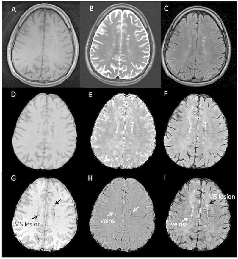
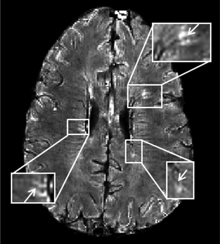
Gradient Echo Plural Contrast Imaging (GEPCI), a fast MRI technique producing inherently co-registered images for qualitative and quantitative assessment of MS, was used to 1) correlate with disability; 2) distinguish clinical MS subtypes; 3) determine prevalence of veins co-localized within lesions in WM.
Thirty subjects representing relapsing-remitting MS (RRMS), secondary progressive MS (SPMS) and primary progressive MS (PPMS) subtypes were scanned with clinical and GEPCI protocols. Standard measures of physical disability and cognition were correlated with magnetic resonance metrics. Lesions with central veins were counted for RRMS subjects.
Tissue damage load (TDL-GEPCI) and lesion load (LL-GEPCI) derived with GEPCI correlated better with MS functional composite (MSFC) measures and most other neurologic measures than lesion load derived with FLAIR (LL-FLAIR). GEPCI correctly classified clinical subtypes in 70% subjects. A central vein could be identified in 76% of WM lesions in RRMS subjects on GEPCI T2*-SWI images.
GEPCI lesion metrics correlated better with neurologic disability than lesion load derived using FLAIR imaging, and showed promise in classifying clinical subtypes of MS. These improvements are likely attributable to the ability of GEPCI to quantify tissue damage.
传统磁共振成像(MRI)方法无法定量多发性硬化症(MS)白质病变的严重程度,也无法测量正常外观白质(NAWM)内的病变。
梯度回波多重对比成像(GEPCI)是一种快速 MRI 技术,可对 MS 进行定性和定量评估,产生固有配准的图像,用于:1)与残疾相关联;2)区分临床 MS 亚型;3)确定 WM 内病变内静脉共存的发生率。
30 名受试者分别代表复发缓解型 MS(RRMS)、继发进展型 MS(SPMS)和原发进展型 MS(PPMS)亚型,接受临床和 GEPCI 方案扫描。身体残疾和认知的标准测量与磁共振指标相关联。对 RRMS 受试者的病变内中央静脉进行计数。
GEPCI 衍生的组织损伤负荷(TDL-GEPCI)和病变负荷(LL-GEPCI)与 MS 功能综合评分(MSFC)和大多数其他神经学测量指标的相关性优于 FLAIR 衍生的病变负荷(LL-FLAIR)。GEPCI 能够在 70%的受试者中正确分类临床亚型。在 RRMS 受试者的 GEPCI T2*-SWI 图像上,可以在 76%的 WM 病变中识别中央静脉。
GEPCI 病变指标与神经残疾的相关性优于使用 FLAIR 成像得出的病变负荷,并且在分类 MS 的临床亚型方面显示出潜力。这些改进可能归因于 GEPCI 定量组织损伤的能力。